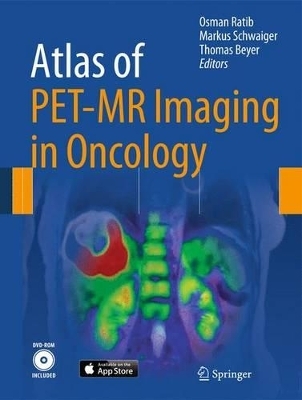
Atlas of PET/MR Imaging in Oncology
- Titel ist leider vergriffen;
keine Neuauflage - Artikel merken
The book opens with an introduction to the principles of hybrid imaging that pays particular attention to PET/MR imaging and standard PET/MR acquisition protocols. A wide range of illustrated clinical case reports are then presented. Each case study includes a short clinical history, findings, and teaching points, followed by illustrations, legends, and comments.
The multimedia version of the book includes dynamic movies that allow the reader to browse through series of rotating 3D images (MIP or volume rendered), display blending between PET and MR, and dynamic visualization of 3D image volumes. The movies can be played either continuously or sequentially for better exploration of sets of images.
The editors of this state-of-the-art publication are key opinion leaders in the field of multimodality imaging. Professor Osman Ratib (Geneva) and Professor Markus Schwaiger (Munich) were the first in Europe to initiate the clinical adoption of PET/MR imaging. Professor Thomas Beyer (Zurich) is an internationally renowned pioneering physicist in the field of hybrid imaging. Individual clinical cases presented in this book are co-authored by leading international radiologists and nuclear physicians experts in the use of PET and MRI.
Osman Ratib is a Professor in Medicine, Chairman of the Department of Medical Imaging and Head of the Division of Nuclear Medicine at the University Hospital of Geneva. Dr. Ratib is board certified in Cardiology and Nuclear Medicine who obtained his medical degrees at the University of Geneva and a Ph.D. in Medical Imaging from University of California Los Angeles in 1989. He was appointed as Professor and Vice Chairman of the Department of Radiology at UCLA from 1998 to 2005 when he returned to Geneva to be appointed as chair of the department of medical imaging and head of the division of Nuclear medicine. In his current position he is responsible of six clinical divisions including radiology, neuroradiology, radio-oncology, nuclear medicine and medical informatics as well as a cyclotron and pre-clinical imaging unit. He has pioneered several innovative projects including advanced cardiovascular PET/CT program and the first whole-body PET/MRI unit in Europe. He is also founding member and president of the OsiriX foundation, a non-profit organization for the promotion of Open-Source software in medicine.
Introduction.- Hybrid imaging principles: From PET/CT to PET/MR.- Technical principles and protocols of PET/MR imaging.- PET/ MR atlas of clinical cases in oncology: Head and neck cancers.- Prostate cancers.- Breast cancers.- Gynecological cancers.- Brain tumors.- Pediatric oncology.- Lymphoma, lung and other tumors.-. Thyxroid and ednocrine tumors.- Benign, generative and inflammatory disease.
| Erscheint lt. Verlag | 28.6.2013 |
|---|---|
| Zusatzinfo | VIII, 232 p. 351 illus., 250 illus. in color. With DVD. |
| Verlagsort | Berlin |
| Sprache | englisch |
| Maße | 210 x 279 mm |
| Gewicht | 889 g |
| Themenwelt | Medizin / Pharmazie ► Medizinische Fachgebiete ► Onkologie |
| Medizinische Fachgebiete ► Radiologie / Bildgebende Verfahren ► Nuklearmedizin | |
| Medizinische Fachgebiete ► Radiologie / Bildgebende Verfahren ► Radiologie | |
| Schlagworte | Atlas • MR • Oncology • Onkologie • PET • Positronen-Emissions-Tomographie (PET) |
| ISBN-10 | 3-642-31291-8 / 3642312918 |
| ISBN-13 | 978-3-642-31291-5 / 9783642312915 |
| Zustand | Neuware |
| Haben Sie eine Frage zum Produkt? |

