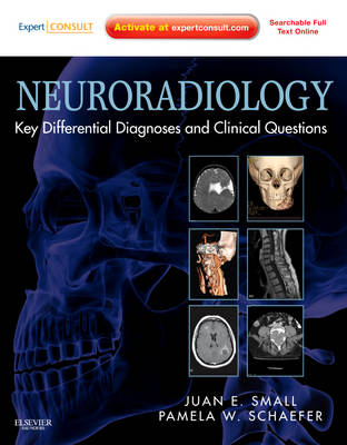
Neuroradiology: Key Differential Diagnoses and Clinical Questions
Expert Consult - Online and Print
Seiten
2012
Saunders (Verlag)
978-1-4377-1721-1 (ISBN)
Saunders (Verlag)
978-1-4377-1721-1 (ISBN)
- Titel erscheint in neuer Auflage
- Artikel merken
Zu diesem Artikel existiert eine Nachauflage
Draws upon Massachusetts General Hospital's vast case collection to help you master the skills you need for interpreting imaging of the head, neck, brain, and spine. This book equips you to make efficient, accurate diagnoses and prepare for imaging exams with hundreds of high-quality, unknown cases in neuroradiology.
"Neuroradiology: Key Differential Diagnoses and Clinical Questions" equips you to make efficient, accurate diagnoses and prepare for imaging exams with hundreds of high-quality, unknown cases in neuroradiology. Drs. Juan Small and Pamela Schaefer draw upon Massachusetts General Hospital's vast case collection to help you master the skills you need for interpreting imaging of the head, neck, brain, and spine.
Key Features:
- Apply systematic pattern analysis techniques to distinguish similar-looking pathological entities from one another.
- Avoid diagnostic pitfalls by recognizing significant variations in the clinical presentation of various diseases.
- See how diagnostic ambiguities are resolved by viewing corresponding gross pathologic and histologic images.
- Benefit from the volume and exceptional quality of Massachusetts General Hospital's patient case load.
- Access the full text and images online, perform rapid searches, and more at expertconsult.com.
Juan Small, MD and Pamela W Schaefer, MD, Associate Professor of Radiology, Massachusetts General Hospital, Boston, Massachusetts
PART 1 BRAIN AND COVERINGS
- Case 1 Computed Tomography Hyperdense Lesions, 3
- Case 2 T1 Hyperintense Lesions, 7
- Case 3 Multiple Susceptibility Artifact Lesions, 13
- Case 4 Ring-Enhancing Lesions, 19
- Case 5 Leptomeningeal Enhancement, 25
- Case 6 Dural Enhancement, 29
- Case 7 Lesions Containing Fat, 35
- Case 8 Extraaxial Lesions, 43
- Case 9 Bilateral Central Gray Matter Abnormality, 53
- Case 10 Temporal Lobe Lesions, 63
- Case 11 Temporal Lobe Cystic Lesions, 67
- Case 12 Cerebellopontine Angle Cisterns, 73
- Case 13 Lateral Ventricular Lesions, 81
- Case 14 Third Ventricular Lesions, 87
- Case 15 Fourth Ventricular Lesions, 93
- Case 16 Suprasellar Cystic Lesions, 99
- Case 17 Pineal Region, 107
- Case 18 Cranial Nerve Lesions, 113
- Case 19 Lytic Skull Lesions, 119
- Case 20 Skull Fracture Versus Sutures, 125
- Case 21 Clivus Lesions, 129
- Case 22 Hyperdense Cerebellum, 135
- Case 23 T2-Hyperintense Pontine Abnormalities, 141
- Case 24 Cerebral Cortical Neurodegeneration, 147
- Case 25 Cerebral Subcortical Neurodegeneration, 155
- Case 26 Epidermoid Versus Arachnoid Cyst, 163
- Case 27 Cyst With A Mural Nodule, 167
- Case 28 Ecchordosis Physaliphora Versus Chordoma, 171
- Case 29 Atlantooccipital And Atlantoaxial Separation, 179
- Case 30 Basilar Invagination And Platybasia, 187
- Case 31 Enhancing Intramedullary Spinal Cord Lesions, 195
- Case 32 Enhancing Intramedullary Conus Lesions, 207
- Case 33 Solitary Enhancing Intradural, Extramedullary Lesions, 211
- Case 34 Multiple Enhancing Intradural, Extramedullary Lesions, 219
- Case 35 Cystic Intradural Extramedullary Lesions, 225
- Case 36 Nerve Root Enlargement, 233
- Case 37 Posterior Element Lesions, 239
- Case 38 Sacral Masses, 243
- Case 39 Disk Infection Versus Inflammatory/Degenerative Changes, 249
- Case 40 Vertebral Compression Fractures, 253
- Case 41 Periauricular Cystic Lesions, 259
- Case 42 Cystic Lateral Neck Masses, 269
- Case 43 Infrahyoid Neck Cystic Lesions, 277
- Case 44 Prestyloid Parapharyngeal Space, 285
- Case 45 Post Styloid Parapharyngeal Space, 289
- Case 46 Floor Of Mouth, 293
- Case 47 Thyroglossal Duct Abnormalities, 297
- Case 48 Anterior Skull Base Masses, 301
- Case 49 Petrous Apex, 309
- Case 50 External Auditory Canal, 315
- Case 51 Middle Ear, 321
- Case 52 Lesions Of The Facial Nerve, 325
- Case 53 Lytic/Cystic Mandibular Lesions, 329
- Case 54 Jugular Foramen Lesions, 333
- Case 55 Optic Nerve Mass, 339
- Case 56 Dilated Superior Ophthalmic Vein/Asymmetric Cavernous Sinus Enhancement, 343
- Case 57 Lacrimal Gland, 347
- Case 58 Nasal Cavity Lesions, 353
- Case 59 Solitary Parotid Mass, 359
- Case 60 Bilateral Parotid Masses, 363
- Case 61 Retropharyngeal Space, 369
- Case 62 Intraventricular Posterior Fossa Tumors, 381
- Case 63 Pediatric Cerebellar Tumors, 385
- Case 64 Pediatric Extraaxial Posterior Fossa Tumors, 389
- Case 65 Midline Posterior Fossa Extraaxial Cystic Lesions, 393
- Case 66 Occipital Cephalocele, 399
- Case 67 Holoprosencephaly, 403
- Case 68 Leukodystrophies, 411
- Case 69 Congenital Arterial Anastamosis, 417
- Case 70 Odontoid: Acute Versus Chronic, 421
- Case 71 Pediatric Nasofrontal Mass, 425
- Case 72 Pediatric Globe Lesions, 429
| Zusatzinfo | Approx. 2000 illustrations (75 in full color) |
|---|---|
| Verlagsort | Philadelphia |
| Sprache | englisch |
| Maße | 190 x 265 mm |
| Gewicht | 1338 g |
| Einbandart | gebunden |
| Themenwelt | Medizin / Pharmazie ► Medizinische Fachgebiete ► Chirurgie |
| Medizin / Pharmazie ► Medizinische Fachgebiete ► Neurologie | |
| Medizin / Pharmazie ► Medizinische Fachgebiete ► Pädiatrie | |
| Medizinische Fachgebiete ► Radiologie / Bildgebende Verfahren ► Radiologie | |
| ISBN-10 | 1-4377-1721-7 / 1437717217 |
| ISBN-13 | 978-1-4377-1721-1 / 9781437717211 |
| Zustand | Neuware |
| Haben Sie eine Frage zum Produkt? |
Mehr entdecken
aus dem Bereich
aus dem Bereich
Buch (2023)
Thieme (Verlag)
190,00 €


