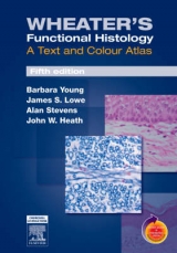
Wheater's Functional Histology
A Text and Colour Atlas
Seiten
2000
|
4th Revised edition
Churchill Livingstone (Verlag)
978-0-443-05612-3 (ISBN)
Churchill Livingstone (Verlag)
978-0-443-05612-3 (ISBN)
- Titel erscheint in neuer Auflage
- Artikel merken
Zu diesem Artikel existiert eine Nachauflage
Presents the photomicrographs and electron micrographs of human tissues along with a concise text for medical and other bioscience courses. This work examines the basic tissue types of blood, supporting/connective tissue, epithelial tissue, muscle and nervous tissues. It also details the microstructure of major body systems.
"Wheater's Functional Histology" presents the student with a resource of high quality photomicrographs and electron micrographs of (largely) human tissues along with a concise text appropriate for modern medical and other bioscience courses. The microstructure of tissues is related to function and clinical significance. The book starts with a section on general cell structure and replication. The second section examines the basic tissue types of blood, supporting/connective tissue, epithelial tissue, muscle and nervous tissues. The third and major section examines the microstructure of the major body systems. For each chapter there is an introductory description followed by a collection of illustrations, each with an extended explanatory caption. This book is ideal for those courses where considerable time is still devoted to the subject. "Functional Histology" will be used as an atlas to go alongside one of the major discursive textbooks. For shorter courses "Functional Histology" will be used as the sole text. The great strength of this book is that it is written from the point of view of the student.
Each chapter has a brief overview of the topic, but most of the information is in the explanatory captions that accompany the high quality photomicrographs.
Cell structure and function Cell cycle and replication Blood Supporting/connective tissues Epithelial tissues Muscle Nervous tissues Circulatory system Skin Skeletal tissues Immune system Respiratory system Oral tissues Gastrointestinal tract Liver and pancreas Urinary system The endocrine glands Male reproductive system Female reproductive system Central nervous system Special sense organs
| Erscheint lt. Verlag | 20.3.2000 |
|---|---|
| Zusatzinfo | 815 ills. |
| Verlagsort | London |
| Sprache | englisch |
| Maße | 203 x 279 mm |
| Gewicht | 1580 g |
| Themenwelt | Studium ► 1. Studienabschnitt (Vorklinik) ► Histologie / Embryologie |
| ISBN-10 | 0-443-05612-9 / 0443056129 |
| ISBN-13 | 978-0-443-05612-3 / 9780443056123 |
| Zustand | Neuware |
| Haben Sie eine Frage zum Produkt? |
Mehr entdecken
aus dem Bereich
aus dem Bereich
Zytologie, Histologie und mikroskopische Anatomie
Buch | Hardcover (2022)
Urban & Fischer in Elsevier (Verlag)
54,00 €
Gewebelehre, Organlehre
Buch | Spiralbindung (2024)
Urban & Fischer in Elsevier (Verlag)
25,00 €
