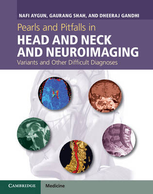
Pearls and Pitfalls in Head and Neck and Neuroimaging
Cambridge University Press (Verlag)
978-1-107-02664-3 (ISBN)
- Titel ist leider vergriffen;
keine Neuauflage - Artikel merken
Nafi Aygun is Associate Professor of Radiology and Director of the Neuroradiology Fellowship Program, Johns Hopkins University, Baltimore, MD, USA. Gaurang Shah is Associate Professor of Radiology, University of Michigan Health System, Ann Arbor, MI, USA. Dheeraj Gandhi is Professor of Radiology, Neurology and Neurosurgery at the University of Maryland School of Medicine, Baltimore, MD, USA.
Part I. Cerebrovascular Diseases; Part II. Demyelinating and Inflammatory Diseases; Part III. Tumors; Part IV. Infectious Diseases; Part V. Metabolic and Neurodegenerative; Part VI. Trauma; Part VII. Miscellaneous; Part VIII. Artifacts and Anatomic Variations; Part IX. Skull Base; Part X. T-Bone; Part XI. Paranasal Sinuses; Part XII. Orbits; Part XIII. Salivary Glands; Part XIV. Neck; Part XV. Thyroid Parathyroid; Part XVI. Vessels; Part XVII. Spinal Column; Part XVIII. Intervertebral Discs.
| Erscheint lt. Verlag | 5.12.2013 |
|---|---|
| Zusatzinfo | 18 Halftones, color; 104 Halftones, black and white; 2 Line drawings, black and white |
| Verlagsort | Cambridge |
| Sprache | englisch |
| Maße | 223 x 283 mm |
| Gewicht | 2000 g |
| Themenwelt | Medizinische Fachgebiete ► Radiologie / Bildgebende Verfahren ► Kernspintomographie (MRT) |
| Medizinische Fachgebiete ► Radiologie / Bildgebende Verfahren ► Radiologie | |
| ISBN-10 | 1-107-02664-4 / 1107026644 |
| ISBN-13 | 978-1-107-02664-3 / 9781107026643 |
| Zustand | Neuware |
| Haben Sie eine Frage zum Produkt? |
aus dem Bereich


