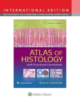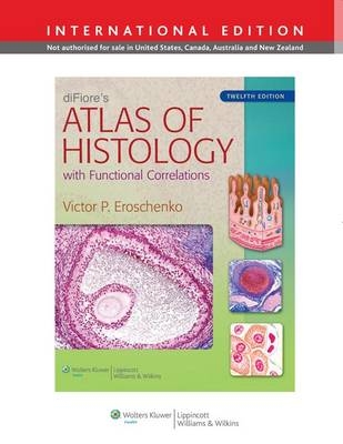
diFiore's Atlas of Histology with Functional Correlations
Seiten
2012
|
Twelfth, International Edition
Lippincott Williams and Wilkins (Verlag)
978-1-4511-7561-5 (ISBN)
Lippincott Williams and Wilkins (Verlag)
978-1-4511-7561-5 (ISBN)
- Titel erscheint in neuer Auflage
- Artikel merken
Zu diesem Artikel existiert eine Nachauflage
Explains basic histology concepts through realistic, full-color composite and idealized illustrations of histologic structures. This title offers an introduction on basic histology techniques and staining as well as a more comprehensive list of stains that students may encounter in their histology course.
diFiore's Atlas of Histology with Functional Correlations explains basic histology concepts through realistic, full-color composite and idealized illustrations of histologic structures. Added to the illustrations are actual photomicrographs of similar structures, a popular trademark of the atlas. All structures are directly correlated with the most important and essential functional correlations, allowing students to efficiently learn histologic structures and their major functions at the same time.
This new edition features:"
Expanded Introduction on basic histology techniques and staining as well as a more comprehensive list of stains that students may encounter in their histology course
· New chapter on cell biology accompanied by both drawings and representative photomicrographs of the main stages in the cell cycle during mitosis
· Contents reorganized into four parts, progressing logically from Methods and Microscopy through Tissues and Systems"
Improved art program with digitally enhanced images to provide increased detailMore than 40 new photomicrograph images, including light and transmission electron micrographs
Student Resources: Online E-book, Interactive Question Bank for chapter review, and Interactive Atlas featuring all images from the book + more than 450 additional micrographs
diFiore’s Atlas of Histology is the perfect resource for medical and graduate histology students.
| Zusatzinfo | 385 |
|---|---|
| Verlagsort | Philadelphia |
| Sprache | englisch |
| Maße | 213 x 276 mm |
| Gewicht | 1247 g |
| Themenwelt | Studium ► 1. Studienabschnitt (Vorklinik) ► Histologie / Embryologie |
| ISBN-10 | 1-4511-7561-2 / 1451175612 |
| ISBN-13 | 978-1-4511-7561-5 / 9781451175615 |
| Zustand | Neuware |
| Haben Sie eine Frage zum Produkt? |
Mehr entdecken
aus dem Bereich
aus dem Bereich
Zytologie, Histologie und mikroskopische Anatomie
Buch | Hardcover (2022)
Urban & Fischer in Elsevier (Verlag)
54,00 €
Gewebelehre, Organlehre
Buch | Spiralbindung (2024)
Urban & Fischer in Elsevier (Verlag)
25,00 €
