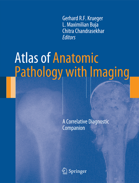
Atlas of Anatomic Pathology with Imaging
A Correlative Diagnostic Companion
Seiten
2013
Springer London Ltd (Verlag)
978-1-4471-2845-8 (ISBN)
Springer London Ltd (Verlag)
978-1-4471-2845-8 (ISBN)
Atlas of Anatomic Pathology with Imaging - A Correlative Diagnostic Companion is a valuable teaching tool for medical students and residents in several specialities such as pathology, radiology, internal medicine, surgery and neurologic sciences. Its need is all the more urgent given the severe shortcuts in the teaching of anatomic pathology following the decrease in the number of autopsies performed. Many of the images shown in the atlas would not be available without performing autopsies and therefore this atlas is an essential for all those in the field.
Atlas of Anatomic Pathology with Imaging - A Correlative Diagnostic Companion is the first to combine gross anatomic pictures of diseases with diagnostic imaging. This unique collection of material consisting of over 2000 illustrations complied by experts from around the world is a valuable diagnostic resource for all medical professionals.
Atlas of Anatomic Pathology with Imaging - A Correlative Diagnostic Companion is the first to combine gross anatomic pictures of diseases with diagnostic imaging. This unique collection of material consisting of over 2000 illustrations complied by experts from around the world is a valuable diagnostic resource for all medical professionals.
Introduction.- Cardiovascular Pathology.- Pathology of the Respiratory Tract.- Pathology of the Gastrointestinal Tract.- Pathology of the Liver, Biliary System, and Exocrine Pancreas.- Pathology of the Urinary System and the Male Genital Tract.- Pathology of the Breast and Female Genital Tract.- Pathology of Hematopoietic and Lymphatic Tissues.- Pathology of Bones and Soft Tissues.- Pathology of Skin and Adnexa.- Endocrine Pathology.- Pathology of the Ear, Nose, and Throat.- Dental and Orofacial Pathology.- Pathology of the Central and Peripheral Nervous System.- Pathology of the Eye.
| Mitarbeit |
Anpassung von: Chitra Chandrasekhar |
|---|---|
| Zusatzinfo | XIV, 821 p. |
| Verlagsort | England |
| Sprache | englisch |
| Maße | 210 x 279 mm |
| Themenwelt | Medizinische Fachgebiete ► Radiologie / Bildgebende Verfahren ► Radiologie |
| Studium ► 1. Studienabschnitt (Vorklinik) ► Anatomie / Neuroanatomie | |
| Studium ► 2. Studienabschnitt (Klinik) ► Pathologie | |
| Schlagworte | anatomy • Microscopic photos • Pathology |
| ISBN-10 | 1-4471-2845-1 / 1447128451 |
| ISBN-13 | 978-1-4471-2845-8 / 9781447128458 |
| Zustand | Neuware |
| Haben Sie eine Frage zum Produkt? |
Mehr entdecken
aus dem Bereich
aus dem Bereich
Buch (2023)
Thieme (Verlag)
190,00 €


