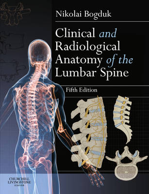
Clinical and Radiological Anatomy of the Lumbar Spine
Churchill Livingstone (Verlag)
978-0-7020-4342-0 (ISBN)
- Titel erscheint in neuer Auflage
- Artikel merken
Clinical and Radiological Anatomy of the Lumbar Spine 5e continues to offer practical, comprehensive coverage of the subject area in a unique single volume which successfully bridges the gap between the basic science of the lumbar region and findings commonly seen in the clinic.
Prepared by an author of international renown, Clinical and Radiological Anatomy of the Lumbar Spine 5e provides clear anatomical descriptions of the individual components of the lumbar region, as well as the intact spine, accompanied by a full colour artwork programme. Detailed anatomical descriptions are followed by an explanation of the basic principles of biomechanics and spinal movement together with a comprehensive overview of embryology and the influence of age-related change in the lumbar region. The problem of low back pain and instability are also fully explored while an expanded section on medical imaging completes the volume.
Clinical and Radiological Anatomy of the Lumbar Spine 5e offers practical, validated and clinically relevant information to all practitioners and therapists working in the field of low back pain and will be ideal for students and practitioners of chiropractic, osteopathic medicine and osteopathy, physiotherapy, physical therapy, pain medicine and physiatry worldwide.
Presents a clear and accessible overview of the basic science relating to the structure and function of the lumbar spine
Written by an internationally renowned expert in the fields of both clinical anatomy and back pain
Describes the structure of the individual components of the lumbar spine, as well as the intact spine
Goes beyond the scope of most anatomy books by endeavouring to explain why the vertebrae and their components are constructed the way they are
Provides an introduction to biomechanics and spinal movement with special emphasis on the role of the lumbar musculature
Explores both embryology and the process of aging in the context of spinal structure and function
Explores mechanical back pain within the context of the structural and biomechanical principles developed earlier in the volume
Extensive reference list allows readers seeking to undertake research projects on some aspect of the lumbar spine with a suitable starting point in their search through the literature
Perfect for use both as an initial resource in undergraduate training in physiotherapy and physical medicine or as essential reading for postgraduate studies
Greatly expanded section on medical imaging
Increased elaboration of the regional anatomy of the lumbar spine
Includes chapter on reconstructive anatomy, which provides an algorithm showing how to put the lumbar spine back together
Presents an ethos of 'anatomy by expectation' - to show readers what to expect on an image, rather than being required to identify what is seen
I commenced research into spinal pain, in 1972, when essentially nothing was known about the problem. There being no established groups or departments working on this problem, I forged my own career, using borrowed resources. I commenced in a Department of Anatomy, where I pursued the innervation of the vertebral column as a fundamental element in understanding the sources and mechanisms of spinal pain. Professor Jim Lance fostered this interest, and accommodated my PhD studies. In his department I continued my anatomy studies but was able also to commence clinical applications. I developed and tested new diagnostic and surgical procedures for back pain and for neck pain. While in Professor Lance's Department, I participated in laboratory studies of the mechanisms of migraine. At the University of Queensland I continued to develop and apply the diagnostic and surgical techniques that I started at the University of NSW, serving as an honorary medical officer at the Pain Clinic of Princess Alexandra Hospital. Meanwhile I supervised science and medicine postgraduate students who undertook basic science studies into the biomechanics of the back and neck. At the University of Newcastle, I had established a reputation sufficient to attract a grant from the Motor Accidents Authority of NSW to investigate the cause and treatment of neck pain after whiplash. The grant supported three PhD students over a six year period. They performed studies that validated the diagnostic procedures and which tested the surgical procedures in a placebo-controlled double-blind randomized trial. Having established an international standing in the development and testing of treatments for spinal pain, I participated in the design and analysis of controlled trials conducted elsewhere in Australia and in the USA. These tested the efficacy of: lumbar radiofrequency neurotomy for back pain, intradiscal electrothermal anuloplasty for back pain, prolotherapy for back pain, exercises for neck pain. Between 1997 and 2002 I conducted the National Musculoskeletal Medicine Initiative which developed and tested evidence-based practice guidelines for the management of back pain, neck pain, shoulder pain, knee pain, and pain in the foot, wrist, and elbow. My work has been awarded the Volvo Award for Back Pain Research, the Research Prize of the Cervical Spine Research Society, the Award for Outstanding Research of the North American Spine Society, and three times the Research Prize of the Spine Society of Australia. My students have been awarded research prizes by the International Association for the Study of Pain, the Australian Rheumatology Association, and the Australian New Zealand College of Anaesthetists. I have never had a funded department to which to attract investigators and academics. I have relied on scholarships for students, and the goodwill of private practitioners who wished to contribute to clinical research. Of late, I have been supervising Neurosurgery residents undertaking studies of the outcomes of treatment for Radicular pain and back pain.
1. The lumbar vertebrae
2. The interbody joint and the intervertebral discs
3. The zygapophysial joints - detailed structure
4. The ligaments of the lumbar spine
5. The lumbar lordosis and the vertebral canal
6. The sacrum
7. Basic biomechanics
8. Movements of the lumbar spine
9. The lumbar muscles and their fasciae
10. Nerves of the lumbar spine
11. Blood supply of the lumbar spine
12. Embryology and development
13. Age changes in the lumbar spine
14. The sacroiliac joint
15. Low back pain
16. Instability
17. Reconstructive anatomy
18. Radiographic anatomy
19. Sagittal magnetic resonance scans
20. Axial magnetic resonance imaging
Appendix
| Zusatzinfo | Approx. 538 illustrations (384 in full color); Illustrations |
|---|---|
| Verlagsort | London |
| Sprache | englisch |
| Maße | 189 x 246 mm |
| Gewicht | 619 g |
| Themenwelt | Medizin / Pharmazie ► Physiotherapie / Ergotherapie |
| Studium ► 1. Studienabschnitt (Vorklinik) ► Anatomie / Neuroanatomie | |
| ISBN-10 | 0-7020-4342-7 / 0702043427 |
| ISBN-13 | 978-0-7020-4342-0 / 9780702043420 |
| Zustand | Neuware |
| Haben Sie eine Frage zum Produkt? |
aus dem Bereich


