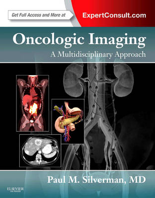
Oncologic Imaging: A Multidisciplinary Approach
W B Saunders Co Ltd (Verlag)
978-1-4377-2232-1 (ISBN)
- Titel erscheint in neuer Auflage
- Artikel merken
Here's the multidisciplinary guidance you need for optimal imaging of malignancies. Radiologists, surgeons, medical oncologists, and radiation oncologists offer state-of-the-art guidelines for diagnosis, staging, and surveillance, equipping all members of the cancer team to make the best possible use of today's noninvasive diagnostic tools.
Consult with the best. Dr. Paul M. Silverman and more than 100 other experts from MD Anderson Cancer Center provide you with today's most dependable answers on every aspect of the diagnosis, treatment, and management of the cancer patient.
Recognize the characteristic presentation of each cancer via current imaging modalities and understand the clinical implications of your findings.
Effectively use traditional imaging modalities such as Multidetector CT (MDCT), PET/CT, and MR in conjunction with the latest advances in molecular oncology and targeted therapies.
Find information quickly and easily thanks to a consistent, highly templated format complete with "Key Point" summaries, algorithms, drawings, and full-color staging diagrams.
Make confident decisions with guidance from comprehensive algorithms for better staging and imaging evaluation.
Access the fully searchable text online, along with high-quality downloadable images for use in teaching and lecturing and online-only algorithms, at expertconsult.com.
Oncologic Imaging Table of Contents
Introduction
Part 1: General Principles
1. A Multidisciplinary Approach to Cancer: A Radiologist's View
2. A Multidisciplinary Approach to Cancer: A Surgeon's View
3. A Multidisciplinary Approach to Cancer: A Medical Oncologist's View
4. A Multidisciplinary Approach to Cancer: A Radiation Oncologist's View
5. Assessing Response to Therapy
Part 2: Chest
6. Lung Cancer
7. Primary Mediastinal Neoplasms
8. Pleural Tumors
Part 3: Liver, Biliary Tract and Pancreas
9. Liver Cancer: Hepatocellular And Fibrolamellar Carcinoma
10. Cholangiocarcinoma
11. Pancreatic Ductal Adenocarcinoma
12. Cystic Pancreatic Lesions
13. Pancreatic Neuroendocrine Tumors
Part 4: Gastrointestinal Tract
14. Esophageal Cancer
15. Gastric Carcinoma
16. Small Bowel Malignant Tumors
17. Colorectal Cancer
Part 5: Genitourinary
18. Renal Tumors
19. Bladder Cancer and Upper Tracts
20. Testicular Germ Cell Tumors
21. Primary Adrenal Malignancy
22. Prostate Cancer
23. Primary Retroperitoneal Tumors
Part 6: Gynecologic and Women's Imaging
24. Tumors of the Uterine Corpus
25. Cervical Cancer
26. Ovarian Cancer
27. Breast Cancer
Part 7: Lymphomas and Hematological Imaging
28. Myeloma and Leukemia
29. Hematological Malignancy: The Lymphomas
Part 8: Metastatic Disease
30. Thoracic Metastatic Disease
31. Metastases Abdominal-Pelvic Organs
32. Peritoneal Cavity and Gastrointestinal Tract
33. Bone Metastases
34. Cancer of Unknown Primary
Part 9: Miscellaneous
35. Imaging in Thyroid Cancer
36. Melanoma
37. Soft Tissue Sarcomas
Part 10: Complications of Therapy
38. Interventional Imaging in the Oncologic Patient
39. Complications in the Oncologic Patient: Chest
40. Complications in the Oncologic Patient: Abdomen and Pelvis
41. Pulmonary Embolic Disease and Cardiac Tumors
Part 11: Protocols in Oncologic Imaging
42. Protocols for Imaging Studies in the Oncologic Patient
| Zusatzinfo | Approx. 1130 illustrations (150 in full color); Illustrations |
|---|---|
| Verlagsort | London |
| Sprache | englisch |
| Maße | 222 x 281 mm |
| Gewicht | 2676 g |
| Themenwelt | Medizin / Pharmazie ► Medizinische Fachgebiete ► Onkologie |
| Medizin / Pharmazie ► Medizinische Fachgebiete ► Radiologie / Bildgebende Verfahren | |
| ISBN-10 | 1-4377-2232-6 / 1437722326 |
| ISBN-13 | 978-1-4377-2232-1 / 9781437722321 |
| Zustand | Neuware |
| Haben Sie eine Frage zum Produkt? |
aus dem Bereich


