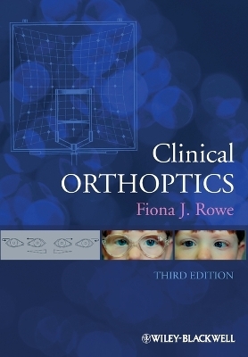
Clinical Orthoptics
Wiley-Blackwell (Verlag)
978-1-4443-3934-5 (ISBN)
FEATURES
Essential reading for students of orthoptics and ophthalmolology
Now fully revised and updated
Generously illustrated with photographs and line drawings
Includes diagnostic aids, case reports, and helpful glossary
Fiona J. Rowe is Lecturer in Orthoptics at the University of Liverpool and an Honorary Research Vision Scientist at the Department of Orthoptics and Ophthalmology, Warrington Hospital. Dr Rowe also lectures extensively to trainee and qualified ophthalmologists, ophthalmic nurses, and other members of the multi-disciplinary eye care team.
Preface xi
Acknowledgements xii
List of Figures xiii
List of Tables xvii
Section I 1
1 Extraocular Muscle Anatomy and Innervation 3
Muscle pulleys 3
Ocular muscles 5
Innervation 10
Associated cranial nerves 12
References 15
Further reading 16
2 Binocular Single Vision 17
Worth’s classification 17
Development 17
Retinal correspondence 19
Physiology of stereopsis 20
Fusion 23
Retinal rivalry 24
Suppression 24
Diplopia 25
References 27
Further reading 28
3 Ocular Motility 29
Saccadic system 29
Smooth pursuit system 31
Vergence system 33
Vestibular-ocular response and optokinetic response 35
Brainstem control 37
Muscle sequelae 39
Past-pointing 40
Bell’s phenomenon 41
References 41
Further reading 43
4 Orthoptic Investigative Procedures 45
Visual acuity 45
Cover test 60
Ocular motility 64
Accommodation and convergence 68
Retinal correspondence 73
Fusion 77
Stereopsis 82
Suppression 89
Synoptophore 91
Aniseikonia 97
Fixation 98
Measurement of deviations 99
Hess charts 105
Field of binocular single vision 108
Uniocular field of vision 110
Measurement of torsion 111
Parks-Helveston three-step test 113
Diplopia charts 113
Bielchowsky phenomenon (dark wedge test) 115
Forced duction test 115
Forced generation test 115
Orthoptic exercises 115
References 119
Further reading 124
Section II 129
5 Heterophoria 131
Classification 131
Aetiology 131
Causes of decompensation 132
Esophoria 132
Exophoria 132
Hyperphoria/hypophoria 133
Alternating hyperphoria 133
Alternating hypophoria 133
Cyclophoria 133
Incomitant heterophoria 133
Hemifield slide 133
Investigation of heterophoria 134
Management 135
References 136
Further reading 137
6 Heterotropia 138
Esotropia 138
Factors necessary for development of binocular single vision 139
Constant esotropia with an accommodative element 140
Constant esotropia without an accommodative element 141
Accommodative esotropia 146
Relating to fixation distance 151
Exotropia 155
Hypertropia 168
Hypotropia 168
Cyclotropia 169
Dissociated vertical deviation 170
Dissociated horizontal deviation 172
Quality of life 173
Pseudostrabismus 174
References 175
Further reading 184
7 Microtropia 189
Terminology 189
Classification 190
Investigation 191
Management 194
References 194
Further reading 195
8 Amblyopia and Visual Impairment 197
Classification 197
Aetiology 197
Investigation 198
Management 199
Eccentric fixation 205
Cerebral visual impairment 205
Delayed visual maturation 206
PHACE syndrome 207
References 207
Further reading 212
9 Aphakia 215
Methods of correction 215
Investigation 215
Problems with unilateral aphakia 216
Management 216
References 218
Further reading 219
Section III 221
10 Incomitant Strabismus 223
Aetiology 223
Aid to diagnosis 225
Diplopia 226
Abnormal head posture 227
References 230
Further reading 231
11 A and V Patterns 232
Classification 232
Aetiology 232
Investigation 236
Management 238
References 241
Further reading 243
12 Accommodation and Convergence Disorders 245
Accommodative disorders 245
Presbyopia – physiological 245
Presbyopia – premature (non-physiological) 246
Accommodative insufficiency 247
Accommodative fatigue 248
Accommodative paralysis 248
Accommodative spasm 249
Accommodative inertia 250
Micropsia 251
Macropsia 251
Convergence anomalies 251
Convergence insufficiency 252
Convergence paralysis 254
Convergence spasm 254
Specific learning difficulty 254
References 255
Further reading 257
13 Ptosis and Pupils 259
Ptosis 259
Marcus Gunn jaw-winking syndrome 263
Lid retraction 264
Pupils 264
References 269
Further reading 271
14 Neurogenic Disorders 272
III (third) cranial nerve 272
IV (fourth) cranial nerve 280
VI (sixth) cranial nerve 288
Multiple sclerosis 292
Acquired motor fusion deficiency 293
Non-accidental injury 294
Premature visual impairment 295
Ophthalmoplegia 296
References 300
Further reading 307
15 Mechanical Paralytic Strabismus 310
Congenital cranial dysinnervation disorders 312
Brown’s syndrome 319
Adherence syndrome 324
Moebius syndrome 325
Strabismus fixus syndrome 327
Thyroid eye disease 327
Orbital injuries 333
Blow-out fracture 334
Soft tissue injury 339
Supraorbital fracture 341
Naso-orbital fracture 341
Zygoma fracture 341
Conjunctival shortening syndrome 342
Retinal detachment 342
Cataract 343
Macular translocation surgery 344
References 344
Further reading 350
16 Myogenic Disorders 354
Thyroid eye disease 354
Chronic progressive external ophthalmoplegia 354
Myasthenia gravis 355
Myotonic dystrophy 358
Ocular myositis 358
Kearns–Sayre ophthalmoplegia 359
References 359
Further reading 361
17 Craniofacial Synostoses 362
Plagiocephaly 362
Brachycephaly 362
Scaphocephaly/dolichocephaly 362
Occipital plagiocephaly 362
Apert’s syndrome 363
Craniofrontonasal dysplasia 363
Crouzon’s syndrome 363
Pfeiffer syndrome 363
Saethre–Chotzen syndrome 364
Unicoronal syndrome 364
General signs and symptoms 364
Ocular signs and symptoms 365
Management 365
References 366
Further reading 367
18 Nystagmus 368
Aetiology 368
Classification 368
Investigation 373
Management 375
References 378
Further reading 380
19 Supranuclear and Internuclear Disorders 382
Saccadic movement disorders 382
Smooth pursuit movement disorders 384
Vergence movement disorders 385
Gaze palsy 386
Optokinetic movement disorders 394
Vestibular movement disorders 395
Brainstem syndromes 395
Skew deviation 397
Ocular tilt reaction 398
Ocular investigation 398
Management options 400
References 401
Further reading 405
Section IV Appendices 407
Diagnostic Aids 409
Abbreviations of Orthoptic Terms 418
Diagrammatic Recording of Ocular Motility 424
Diagrammatic Recording of Nystagmus 426
Glossary 428
Case Reports 441
Index 459
| Erscheint lt. Verlag | 9.3.2012 |
|---|---|
| Verlagsort | Hoboken |
| Sprache | englisch |
| Maße | 173 x 244 mm |
| Gewicht | 943 g |
| Themenwelt | Medizin / Pharmazie ► Medizinische Fachgebiete ► Augenheilkunde |
| ISBN-10 | 1-4443-3934-6 / 1444339346 |
| ISBN-13 | 978-1-4443-3934-5 / 9781444339345 |
| Zustand | Neuware |
| Haben Sie eine Frage zum Produkt? |
aus dem Bereich


