Oral Pathology
Clinical Pathologic Correlations
Seiten
2011
|
6th Revised edition
Saunders (Verlag)
978-1-4557-0262-6 (ISBN)
Saunders (Verlag)
978-1-4557-0262-6 (ISBN)
- Titel ist leider vergriffen;
keine Neuauflage - Artikel merken
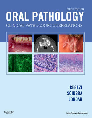
Covering pathologic conditions by clinical appearance, this title uses an atlas-style format to help you identify, diagnose, and plan treatment for oral disease presentations. Each chapter is organized by clinical appearance, and includes full-color photomicrographs and clinical photos to help you identify pathologic elements.
Covering pathologic conditions by clinical appearance, "Oral Pathology: Clinical Pathologic Correlations, 6th Edition" uses an atlas-style format to help you identify, diagnose, and plan treatment for oral disease presentations. Two-page spreads include clinical photos of common conditions on one side while the facing page lists the central features, causes, and significance of each specific disease. Each chapter is organized by clinical appearance, such as white lesions, red-blue lesions, and cysts of the jaws and neck, and includes full-color photomicrographs and clinical photos to help you identify pathologic elements. This edition adds new coverage of oral cancer and new cone beam CT, regular CT, and MRI images. Expert authors Joseph Regezi, James Sciubba, and Richard Jordan provide a quick reference that's ideal for the lab, NBDE review, or chairside use!
Covering pathologic conditions by clinical appearance, "Oral Pathology: Clinical Pathologic Correlations, 6th Edition" uses an atlas-style format to help you identify, diagnose, and plan treatment for oral disease presentations. Two-page spreads include clinical photos of common conditions on one side while the facing page lists the central features, causes, and significance of each specific disease. Each chapter is organized by clinical appearance, such as white lesions, red-blue lesions, and cysts of the jaws and neck, and includes full-color photomicrographs and clinical photos to help you identify pathologic elements. This edition adds new coverage of oral cancer and new cone beam CT, regular CT, and MRI images. Expert authors Joseph Regezi, James Sciubba, and Richard Jordan provide a quick reference that's ideal for the lab, NBDE review, or chairside use!
Clinical Overview 1. Vesiculobullous Diseases 2. Ulcerative Conditions 3. White Lesions 4. Red-Blue Lesions 5. Pigmented Lesions 6. Verrucal-Papillary Lesions 7. Connective Tissue Lesions 8. Salivary Gland Diseases 9. Lymphoid Lesions 10. Cysts of the Jaws and Neck 11. Odontogenic Tumors 12. Benign Nonodontogenic Tumors 13. Inflammatory Jaw Lesions 14. Malignancies of the Jaws 15. Metabolic and Genetic Diseases 16. Abnormalities of Teeth
| Zusatzinfo | Approx. 958 illustrations (769 in full color) |
|---|---|
| Verlagsort | Philadelphia |
| Sprache | englisch |
| Maße | 222 x 281 mm |
| Themenwelt | Medizin / Pharmazie ► Zahnmedizin |
| ISBN-10 | 1-4557-0262-5 / 1455702625 |
| ISBN-13 | 978-1-4557-0262-6 / 9781455702626 |
| Zustand | Neuware |
| Haben Sie eine Frage zum Produkt? |
Mehr entdecken
aus dem Bereich
aus dem Bereich
Buch | Spiralbindung (2023)
Asgard (Verlag)
40,00 €
Lokalanästhesie, Analgesie, Sedierung
Buch | Hardcover (2024)
QUINTESSENZ Verlag
88,00 €
BEL II mit ausführlichem Expertenkommentar sowie Erläuterungen und …
Buch | Spiralbindung (2023)
Spitta GmbH (Verlag)
159,43 €


