


Atlas of Sectional Radiological Anatomy for PET/CT
Seiten
2012
Springer-Verlag New York Inc.
978-1-4614-1526-8 (ISBN)
Springer-Verlag New York Inc.
978-1-4614-1526-8 (ISBN)
The horizons of sophisticated imaging have expanded with the use of combined positron emission tomography (PET) and computed tomography (CT). PET-CT has revolutionized medical imaging by adding anatomic localization to functional imaging, thus providing physicians with information that is vital for the accurate diagnosis and treatment of pathologies. Since the integration of PET and CT several years ago, PET/CT procedures are now routine at leading medical centers throughout the world. This has increased the importance of nuclear medicine physicians acquiring a broad knowledge in sectional anatomy for image interpretation. The Atlas of Sectional Radiological Anatomy for PET/CT is a user-friendly guide presenting high-resolution, full-color images of anatomical detail and focuses solely on normal FDG distribution throughout the head & neck, thorax, abdomen, and pelvis, the primary sites for cancer detection and treatment through PET/CT.
Dr. Mehmet Kitapci is a leading radiologist with Gazi University in Ankara, Turkey.
Head and Neck.- Thorax.- Abdomen.- Pelvis.
| Zusatzinfo | XI, 115 p. |
|---|---|
| Verlagsort | New York, NY |
| Sprache | englisch |
| Maße | 178 x 254 mm |
| Themenwelt | Medizinische Fachgebiete ► Radiologie / Bildgebende Verfahren ► Nuklearmedizin |
| Medizinische Fachgebiete ► Radiologie / Bildgebende Verfahren ► Radiologie | |
| ISBN-10 | 1-4614-1526-8 / 1461415268 |
| ISBN-13 | 978-1-4614-1526-8 / 9781461415268 |
| Zustand | Neuware |
| Haben Sie eine Frage zum Produkt? |
Mehr entdecken
aus dem Bereich

aus dem Bereich
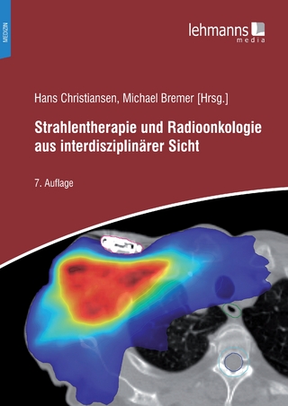
Buch | Softcover (2022)
Lehmanns Media (Verlag)
39,95 €
Lehrbuch für Breast Care Nurses und Fachpersonen in der Onkologie
Buch | Hardcover (2020)
Hogrefe (Verlag)
50,00 €