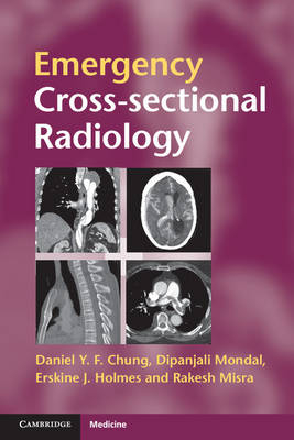
Emergency Cross-sectional Radiology
Seiten
2012
Cambridge University Press (Verlag)
978-0-521-27953-6 (ISBN)
Cambridge University Press (Verlag)
978-0-521-27953-6 (ISBN)
In addition to radiologists, a wide range of clinicians now need to interpret cross-sectional scans of the acutely ill patient. Emergency Cross-sectional Radiology is a highly illustrated rapid reference for emergency medicine physicians, surgeons, acute care physicians, radiologists and any healthcare professional involved in acute care.
Cross-sectional imaging plays an ever-increasing role in the management of the acutely ill patient. There is 24/7 demand for radiologists at all levels of training to interpret complex scans, and alongside this an increased expectation that the requesting physician should be able to recognise important cross-sectional anatomy and pathology in order to expedite patient management. Emergency Cross-sectional Radiology addresses both these expectations. Part I demystifies cross-sectional imaging techniques. Part II describes a wide range of emergency conditions in an easy-to-read bullet point format. High quality images reinforce the findings, making this an invaluable rapid reference in everyday clinical practice. Emergency Cross-sectional Radiology is a practical aide-memoire for emergency medicine physicians, surgeons, acute care physicians and radiologists in everyday reporting or emergency on-call environments.
Cross-sectional imaging plays an ever-increasing role in the management of the acutely ill patient. There is 24/7 demand for radiologists at all levels of training to interpret complex scans, and alongside this an increased expectation that the requesting physician should be able to recognise important cross-sectional anatomy and pathology in order to expedite patient management. Emergency Cross-sectional Radiology addresses both these expectations. Part I demystifies cross-sectional imaging techniques. Part II describes a wide range of emergency conditions in an easy-to-read bullet point format. High quality images reinforce the findings, making this an invaluable rapid reference in everyday clinical practice. Emergency Cross-sectional Radiology is a practical aide-memoire for emergency medicine physicians, surgeons, acute care physicians and radiologists in everyday reporting or emergency on-call environments.
Daniel Y. F. Chung is a Specialist Registrar in Clinical Radiology in the Oxford Deanery and a Clinical Radiology Academic Clinical Fellow at the Oxford University Clinical Academic Graduate School, Oxford. Dipanjali Mondal is a Speciality Registrar in Radiology at the John Radcliffe Hospital, Oxford. Erskine J. Holmes is an Emergency Medicine Consultant at Wexham Park Hospital, Slough. Rakesh Misra is a Consultant Radiologist at Wycombe Hospital, Buckinghamshire.
Preface; Part I. Fundamentals of Cross-sectional Imaging: 1. CT; 2. MRI; 3. Ultrasound; Part II. Pathology: 4. Head; 5. Chest; 6. Abdomen; 7. Musculoskeletal; Index.
| Erscheint lt. Verlag | 19.4.2012 |
|---|---|
| Zusatzinfo | 150 Halftones, unspecified; 50 Line drawings, unspecified |
| Verlagsort | Cambridge |
| Sprache | englisch |
| Maße | 157 x 234 mm |
| Gewicht | 400 g |
| Themenwelt | Medizin / Pharmazie ► Medizinische Fachgebiete ► Notfallmedizin |
| Medizinische Fachgebiete ► Radiologie / Bildgebende Verfahren ► Radiologie | |
| ISBN-10 | 0-521-27953-4 / 0521279534 |
| ISBN-13 | 978-0-521-27953-6 / 9780521279536 |
| Zustand | Neuware |
| Haben Sie eine Frage zum Produkt? |
Mehr entdecken
aus dem Bereich
aus dem Bereich
Buch (2023)
Thieme (Verlag)
190,00 €


