Practical Fluoroscopy of the GI and GU Tracts
Cambridge University Press (Verlag)
978-1-107-00180-0 (ISBN)
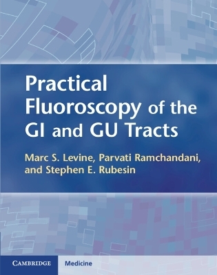
Practical Fluoroscopy of the GI and GU Tracts highlights the critical role of fluoroscopy in the diagnosis of luminal GI and GU diseases, presenting both the fundamentals and nuances for performing and interpreting all types of these examinations. The text presents detailed descriptions of the techniques for performing GI and GU fluoroscopic procedures in a logical, stepwise format. Practical tips, advice and solutions address the problems and pitfalls commonly encountered during these examinations. Clear, concise, yet comprehensive descriptions of the relevant clinical and radiographic findings and differential diagnoses also provide a focused approach for interpreting GI and GU studies. A plethora of carefully annotated figures illustrate the pertinent findings. Practical Fluoroscopy of the GI and GU Tracts is a must-have text for both radiology trainees and experienced radiologists and is an essential addition to the library of every radiology training program and the fluoroscopy suite of every radiology practice.
Marc S. Levine, MD, is Chief of the Gastrointestinal Radiology Section and Professor of Radiology at the University of Pennsylvania Medical Center and Advisory Dean at the University of Pennsylvania School of Medicine, Philadelphia, PA, USA. Parvati Ramchandani, MD, is Chief of the Genitourinary Radiology Section and Professor of Radiology and Surgery at the University of Pennsylvania Medical Center, Philadelphia, PA, USA. Stephen E. Rubesin, MD, is Professor of Radiology at the University of Pennsylvania Medical Center, Philadelphia, PA, USA.
Preface; Part I. GI Tract: 1. Pharynx; 2. Examination of the esophagus, stomach and duodenum: techniques and normal anatomy; 3. Esophagus; 4. Stomach; 5. Duodenum; 6. Small intestine: normal anatomy and techniques; 7. Small intestine; 8. Colon: normal anatomy and techniques; 9. Colon; Part II. GU Tract: 10. Fluoroscopic evaluation of the bladder, urethra and urinary divisions; 11. Retrograde pyelography; Index.
| Erscheint lt. Verlag | 26.1.2012 |
|---|---|
| Zusatzinfo | 600 Halftones, black and white; 2 Line drawings, black and white |
| Verlagsort | Cambridge |
| Sprache | englisch |
| Maße | 225 x 282 mm |
| Gewicht | 1060 g |
| Themenwelt | Medizin / Pharmazie ► Gesundheitsfachberufe ► MTA - Radiologie |
| Medizinische Fachgebiete ► Innere Medizin ► Gastroenterologie | |
| Medizinische Fachgebiete ► Radiologie / Bildgebende Verfahren ► Radiologie | |
| ISBN-10 | 1-107-00180-3 / 1107001803 |
| ISBN-13 | 978-1-107-00180-0 / 9781107001800 |
| Zustand | Neuware |
| Haben Sie eine Frage zum Produkt? |
aus dem Bereich


