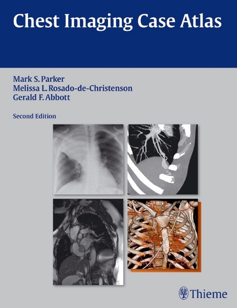
Chest Imaging Case Atlas
Seiten
2012
|
2nd edition
Thieme Medical Publishers Inc (Verlag)
978-1-60406-590-9 (ISBN)
Thieme Medical Publishers Inc (Verlag)
978-1-60406-590-9 (ISBN)
A comprehensive atlas covering the breadth and depth of chest imaging
Written by renowned experts in chest imaging, Chest Imaging Case Atlas, Second Edition enables radiology residents, fellows, and practitioners to hone their diagnostic skills by teaching them how to interpret a large number of radiologic cases. This atlas contains over 200 cases on conditions ranging from Adenoid Cystic Carcinoma to Wegener Granulomatosis. Each case is supported by a discussion of the disease, its underlying pathology, typical and unusual imaging findings, management, and prognosis, providing a comprehensive overview of each disorder.
Special Features of the Second Edition:
Over 1500 high-quality images demonstrating normal and pathologic findings and their variations
More multiplanar, CT angiographic (CTA), MRI, and 3D imaging is incorporated into the text, helping readers stay current with this rapidly changing technology
40 new cases and updated images in cases from the previous edition
A new post-thoracotomy chest section addresses normal post-operative findings and complications associated with common thoracic interventional procedures
The neoplastic diseases section includes the new TNM staging system for lung cancer
The adult cardiovascular disease section now contains a discussion on univentricular and biventricular or end-stage heart failure including various ventricular assist devices and the Total Artificial Heart, their imaging features, and complications associated with their use
The diffuse lung disease section has been expanded to include an approach to HRCT interpretation
Case discussions are based on up-to-datereviews of current literature as well as classic landmark articles
Pearls are provided to describe the features that may strongly support a specific diagnosis, enabling readers to sharpen their clinical diagnostic skills
This book is an invaluable illustrated reference that all physicians in radiology and chest imaging in particular, including pulmonary medicine physicians and thoracic surgeons, should have on their bookshelf.
Associate Professor of Radiology, Harvard University, and Associate Radiologist at Massachusetts General Hospital, Boston, MA, USA
Section I Normal Thoracic Anatomy
Section II Developmental Anomalies
Section III Airways Disease
Section IV Atelectasis
Section V Pulmonary Infection and Aspiration Pneumonia
Section VI Neoplastic Disease
Section VII Thoracic Trauma
Section VIII Diffuse Lung Disease
Section IX Occupational Lung Disease
Section X Adult Cardiovascular Disease
Section XI Abnormalities of the Mediastinum
Section XII Pleura, Chest Wall, and Diaphragm
Section XIII Post-Thoracotomy Chest
| Zusatzinfo | 1584 Illustrations |
|---|---|
| Verlagsort | New York |
| Sprache | englisch |
| Maße | 216 x 279 mm |
| Gewicht | 3266 g |
| Themenwelt | Medizin / Pharmazie ► Gesundheitsfachberufe ► MTA - Radiologie |
| Medizin / Pharmazie ► Medizinische Fachgebiete ► Intensivmedizin | |
| Medizin / Pharmazie ► Medizinische Fachgebiete ► Radiologie / Bildgebende Verfahren | |
| ISBN-10 | 1-60406-590-7 / 1604065907 |
| ISBN-13 | 978-1-60406-590-9 / 9781604065909 |
| Zustand | Neuware |
| Informationen gemäß Produktsicherheitsverordnung (GPSR) | |
| Haben Sie eine Frage zum Produkt? |
Mehr entdecken
aus dem Bereich
aus dem Bereich
Buch | Softcover (2024)
Studia Universitätsverlag Innsbruck
48,00 €
Buch | Hardcover (2024)
Mosby (Verlag)
229,40 €
Buch | Softcover (2024)
Mosby (Verlag)
119,95 €
