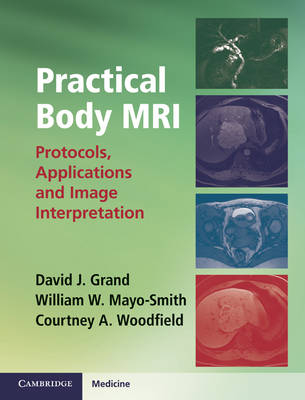
Practical Body MRI
Protocols, Applications and Image Interpretation
Seiten
2012
Cambridge University Press (Verlag)
978-1-107-01404-6 (ISBN)
Cambridge University Press (Verlag)
978-1-107-01404-6 (ISBN)
Practical Body MRI: Protocols, Applications and Image Interpretation demystifies MRI exams of the abdomen and pelvis, explaining everything radiologists should know to appropriately protocol, quality assess and interpret studies. Each chapter illustrates why each sequence is performed, what to look for, and how findings lead to an accurate diagnosis.
Practical Body MRI: Protocols, Applications and Image Interpretation demystifies MRI examinations of the abdomen and pelvis, giving the essential knowledge required by radiologists in order to develop and select appropriate protocols, assess scan quality and interpret imaging studies. Each chapter describes why each sequence is performed, what to look for, and how the important findings from each sequence lead to a unique diagnosis. Numerous protocols are included, from the more common, such as liver and renal MRI, to more tailored examinations such as rectal and placental MRI. All protocols are richly illustrated with images of body MR pathology. A separate chapter discusses MRA/MRV and an introductory chapter gives a brief, practical introduction to MRI physics and receiver coils. The authors' expertise and practical, concise explanations of both protocols and image interpretation makes this an essential resource for residents, fellows and experienced radiologists using body MRI for the first time.
Practical Body MRI: Protocols, Applications and Image Interpretation demystifies MRI examinations of the abdomen and pelvis, giving the essential knowledge required by radiologists in order to develop and select appropriate protocols, assess scan quality and interpret imaging studies. Each chapter describes why each sequence is performed, what to look for, and how the important findings from each sequence lead to a unique diagnosis. Numerous protocols are included, from the more common, such as liver and renal MRI, to more tailored examinations such as rectal and placental MRI. All protocols are richly illustrated with images of body MR pathology. A separate chapter discusses MRA/MRV and an introductory chapter gives a brief, practical introduction to MRI physics and receiver coils. The authors' expertise and practical, concise explanations of both protocols and image interpretation makes this an essential resource for residents, fellows and experienced radiologists using body MRI for the first time.
David J. Grand is Director of Body MRI and Assistant Professor of Diagnostic Imaging at the Warren Alpert Medical School, Brown University, Providence, RI, USA. William W. Mayo-Smith is Director of Computed Tomography and Professor of Diagnostic Radiology at the Warren Alpert Medical School, Brown University, Providence, RI, USA. Courtney A. Woodfield is Staff Radiologist at Diagnostic Imaging Inc., Trevose, PA, USA.
Preface; 1. Introduction to body MRI; 2. NSF guidelines; 3. Liver; 4. MCRP/pancreas; 5. Adrenal/renal; 6. Enterography; 7. Abdomen/pelvis screen; 8. Female pelvis; 9. Pelvic prolapse; 10. Female urethra; 11. Anorectal fistula; 12. Rectal mass; 13. Male pelvis; 14. Pregnant patients; 15. MRA/MRV; Index.
| Erscheint lt. Verlag | 11.10.2012 |
|---|---|
| Zusatzinfo | 50 Tables, black and white; 100 Halftones, unspecified; 100 Line drawings, unspecified |
| Verlagsort | Cambridge |
| Sprache | englisch |
| Maße | 195 x 253 mm |
| Gewicht | 550 g |
| Themenwelt | Medizinische Fachgebiete ► Radiologie / Bildgebende Verfahren ► Kernspintomographie (MRT) |
| ISBN-10 | 1-107-01404-2 / 1107014042 |
| ISBN-13 | 978-1-107-01404-6 / 9781107014046 |
| Zustand | Neuware |
| Haben Sie eine Frage zum Produkt? |
Mehr entdecken
aus dem Bereich
aus dem Bereich
Lehrbuch und Fallsammlung zur MRT des Bewegungsapparates
Buch | Hardcover (2020)
mr-verlag
219,00 €


