Arthritis in Black and White
W B Saunders Co Ltd (Verlag)
978-1-4160-5595-2 (ISBN)
- Titel erscheint in neuer Auflage
- Artikel merken
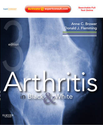
Arthritis in Black and White, by Anne C. Brower, MD and Donald J. Flemming, MD, provides you with a concise, practical introduction to the radiographic diagnosis of arthritic disorders. Completely revised, this popular, easy-to-read resource contains high-quality digital radiographs with correlating MRIs throughout and a practical organization that aids in your recognition, diagnosis, and treatment of common arthritides. In print and online at www.expertconsult.com, it is perfect for residents in training and experienced radiologists wishing to refresh their knowledge.
Easily reference diagnostic guidance by presenting symptom, see what to look for, and understand how to effectively diagnose the patient.
Reference key information quickly and easily thanks to a consistent, user-friendly format and a unique two-part organization (radiologic approaches to specific joints and full description of the individual common arthritides) that facilitates finding the exact information you need for any joint in the body.
Improve the accuracy of your diagnoses by interpreting radiographs and comparing them with correlating MRI images.
Benefit from the latest advancements and techniques found in completely revised and rewritten chapters.
Understand the nuances and subtleties of how arthritides present through over 350 high-quality digital images.
Access the fully searchable text online at www.expertconsult.com, along with a downloadable image bank and more.
1. Imaging Techniques and Modalities
Part I: Approach to Radiographic Changes Observed in a Specific Joint.
2. Evaluation of the Hand Film.
3. Approach to the Foot
4. Approach to the Hip.
5. Approach to the Knee.
6. Approach to the Shoulder.
7. The Sacroiliac Joint.
8. The "Phytes" of the Spine.
Part II: Radiographic Changes Observed in a Specific Articular Disease.
9. Rheumatoid Arthritis.
10. Psoriatic Arthritis.
11. Reactive Arthritis.
12. Ankylosing Spondylitis.
13. Osteoarthritis
14. Neuropathic Osteoarthropaty.
15. Diffuse Isiopathic Skeletal Hyperostosis.
16. Gout.
17. Calcium Pyrophospate Dihydrate Crystal Deposition Disease.
18. Hydroxyapatite Deposition Disease.
19. Miscellaneous Deposition Diseases.
20. Collagen Vascular Diseases (Connective Tissue Diseases).
21. Juvenile Idiopathic Arthritis.
22. Hemophilia.
23. Mass-like Arthropathies.
| Zusatzinfo | 659 illustrations; Illustrations |
|---|---|
| Verlagsort | London |
| Sprache | englisch |
| Maße | 216 x 276 mm |
| Gewicht | 1220 g |
| Themenwelt | Medizin / Pharmazie ► Gesundheitsfachberufe ► MTA - Radiologie |
| Medizin / Pharmazie ► Medizinische Fachgebiete ► Orthopädie | |
| Medizinische Fachgebiete ► Radiologie / Bildgebende Verfahren ► Radiologie | |
| ISBN-10 | 1-4160-5595-9 / 1416055959 |
| ISBN-13 | 978-1-4160-5595-2 / 9781416055952 |
| Zustand | Neuware |
| Informationen gemäß Produktsicherheitsverordnung (GPSR) | |
| Haben Sie eine Frage zum Produkt? |
aus dem Bereich


