Neuroanatomy
Lippincott Williams and Wilkins (Verlag)
978-1-60831-491-1 (ISBN)
- Titel erscheint in neuer Auflage
- Artikel merken
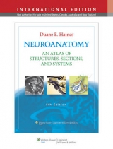
Now in its 25th year, this best-selling work is the only neuroanatomy atlas to integrate neuroanatomy and neurobiology with extensive clinical information. It combines full-color anatomical illustrations with over 200 MRI, CT, MRA, and MRV images to clearly demonstrate anatomical-clinical correlations. This edition contains many new MRI/CT images and is fully updated to conform to Terminologia Anatomica. Fifteen innovative new color illustrations correlate clinical images of lesions at strategic locations on pathways with corresponding deficits in Brown-Sequard syndrome, dystonia, Parkinson disease, and other conditions. The question-and-answer chapter contains over 235 review questions, many USMLE-style. "Interactive Neuroanatomy, Version 3", an online component packaged with the atlas, contains new brain slice series, including coronal, axial, and sagittal slices.
Introduction and Readers Guide Including Rationale for Labels and Abbreviations External Morphology of the Central Nervous System The Spinal Cord: Gross Views and Vasculature The Brain: Lobes, Principle Brodmann Areas, Sensory-Motor Somatotopy The Brain: Gross Views, Vasculature, and MRI The Cerebellum: Gross Views and MRI The Insula: Gross View, Vasculature, and MRI Cranial Nerves Cranial Nerves in MRI Deficits of Eye Movements in the Horizontal Plane Cranial Nerve Deficits in Representative Brainstem Lesions Meninges, Cisterns, Ventricles and Related Hemorrhages The Meninges and Meningeal and Brain Hemorrhages Cisterns and Subarachnoid Hemorrhage Ventricles and Hemorrhage into the Ventricles The Choroid Plexus: Locations, Blood Supply, Tumors Internal Morphology of the Brain in Slices and MRI Brain Slices in the Coronal Plane Correlated with MRI Brain Slices in the Axial Plane Correlated with MRI Internal Morphology of the Spinal Cord and Brain in Stained Sections The Spinal Cord with CT and MRI Arterial Patterns Within the Spinal Cord With Vascular Syndromes The Degenerated Corticospinal Tract The Medulla Oblongata with MRI and CT Arterial Patterns Within the Medulla Oblongata With Vascular Syndromes The Cerebellar Nuclei The Pons with MRI and CT Arterial Patterns Within the Pons with Vascular Syndromes The Midbrain with MRI and CT Arterial Patterns Within the Midbrain With Vascular Syndromes The Diencephalon and Basal Nuclei with MRI Arterial Patterns Within the Forebrain With Vascular Syndromes Internal Morphology of the Brain in Stained Sections: Axial-Sagittal Correlations with MRI Axial-Sagittal Correlations Synopsis of Functional Components, Tracts, Pathways, and Systems: Examples in Anatomical and Clinical Orientation Components of Cranial and Spinal Nerves Orientation Sensory Pathways Motor Pathways Cerebellum and Basal Nuclei Optic, Auditory, and Vestibular Systems Limbic System: Hippocampus and Amygdala Anatomical-Clinical Correlations: Cerebral Angiogram, MRA, and MRV Cerebral Angiogram, MRA, and MRV Overview of Vertebral and Carotid Arteries Questions and Answers: A Sampling of Study and Review Questions, Many in the USMLE Style, All With Explained Answers Sources and Suggested Readings
| Erscheint lt. Verlag | 1.4.2010 |
|---|---|
| Verlagsort | Philadelphia |
| Sprache | englisch |
| Maße | 213 x 277 mm |
| Gewicht | 1112 g |
| Themenwelt | Medizin / Pharmazie ► Gesundheitswesen |
| Medizin / Pharmazie ► Medizinische Fachgebiete ► Neurologie | |
| Medizin / Pharmazie ► Medizinische Fachgebiete ► Psychiatrie / Psychotherapie | |
| Medizin / Pharmazie ► Medizinische Fachgebiete ► Schmerztherapie | |
| Medizin / Pharmazie ► Medizinische Fachgebiete ► Sportmedizin | |
| Medizin / Pharmazie ► Physiotherapie / Ergotherapie ► Rehabilitation | |
| Studium ► 1. Studienabschnitt (Vorklinik) ► Anatomie / Neuroanatomie | |
| ISBN-10 | 1-60831-491-X / 160831491X |
| ISBN-13 | 978-1-60831-491-1 / 9781608314911 |
| Zustand | Neuware |
| Haben Sie eine Frage zum Produkt? |
aus dem Bereich


