Atlas of Cross Sectional Anatomy and Radiological Imaging
Seiten
2012
Anshan Ltd (Verlag)
978-1-84829-057-0 (ISBN)
Anshan Ltd (Verlag)
978-1-84829-057-0 (ISBN)
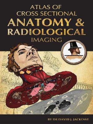
The study of both cadaveric axial cross-sections and CT scans is the basis of 21st century anatomy, and the cornerstone of clinical diagnostics. This text illustrates the sequence of axial cross-sections and CT scans in 3-Dimensional illustrations.
The study of both cadaveric axial cross-sections and CT scans is the basis of 21st century anatomy, and the cornerstone of clinical diagnostics. Modern medical imaging, such as CT (Computed Tomography) scans, produce 1-Dimensional anatomic cross-sections of the axial plane. Learning the proper sequence and orientation of axial cross-sections and CT scans is often extremely challenging, even for the most dedicated students of anatomy: The shapes seen in the axial plane have little relation to the more familiar coronal plane. Most texts abandon students to simply memorize the shapes seen at high-yield vertebral levels or perform tricky mental gymnastics, as they must mentally rotate the axial plane to the more familiar coronal. Students are further frustrated when learning CT scans, as the shapes seen in gray/white CT slices have little relation to the anatomic structures from which they are derived.
This text serves to solve these problems by illustrating the sequence of axial cross-sections and CT scans in unique 3-Dimensional illustrations. This 3-D approach clearly demonstrates the relation of the shapes seen in cross-sections and CT's to their more familiar coronal/sagittal orientation.
The illustrations themselves have been done by Dr Jackowe in the classic style of Vesalius and Bourgery, thus creating a work that is both informative and artistic, the first aesthetic anatomy textbook for many years.
The atlas will serve as a review book, suitable for self-study and as a companion to standard anatomy textbooks. It will appeal to medical/anatomy students, medical residents, and radiologists, as well as the general science reader who will appreciate the quality of the illustrations
I. The Head and Neck - 25 images. Part 1 The Head, Part 2 The Neck
II. The Thorax - 30 images. Part 1 Male Thorax, Part 2, Female Thorax
III. The Abdomen - 20 images.
IV. The Pelvis - 30 images. Part 1. Male Pelvis, Part 2. Female Pelvix
V. The Extremities - 45 images. Part 1. Upper Extremities, Part 2. Lower Extremities.
KEY POINTS
1. 3-Dimensional atlas of cross-sectional anatomy and CT scans.
2. Combines 21st century imaging technology with a classic style of illustration.
3. Novel images of the human body, presented in a unique perspective
4. Color insets with real CT scans superimposed on cross-sections to clearly show the structures behind the scans.
| Erscheint lt. Verlag | 20.9.2012 |
|---|---|
| Zusatzinfo | 100 Illustrations, color |
| Verlagsort | Tunbridge Wells |
| Sprache | englisch |
| Maße | 216 x 278 mm |
| Themenwelt | Studium ► 1. Studienabschnitt (Vorklinik) ► Anatomie / Neuroanatomie |
| ISBN-10 | 1-84829-057-8 / 1848290578 |
| ISBN-13 | 978-1-84829-057-0 / 9781848290570 |
| Zustand | Neuware |
| Informationen gemäß Produktsicherheitsverordnung (GPSR) | |
| Haben Sie eine Frage zum Produkt? |
Mehr entdecken
aus dem Bereich
aus dem Bereich
Struktur und Funktion
Buch | Softcover (2021)
Urban & Fischer in Elsevier (Verlag)
44,00 €
Buch | Hardcover (2022)
Urban & Fischer in Elsevier (Verlag)
220,00 €


