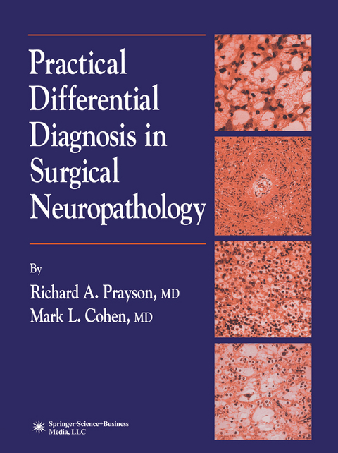
Practical Differential Diagnosis in Surgical Neuropathology - Richard A. Prayson, Mark L. Cohen

Practical Differential Diagnosis in Surgical Neuropathology

Buch | Softcover
179 Seiten
2010 | Softcover reprint of hardcover 1st ed. 2000
Humana Press Inc. (Verlag)
978-1-61737-201-8 (ISBN)
139,09 inkl. MwSt
Not another textbook for neuropathology! Yes, Our publishers helped us with this last problem by we hear you and feel your pain. In fact, that was our insisting on black and white photographs. We ini­ initial response when we were approached to write tially protested, noting that many recent textbook the book you are now holding. In surveying the reviews seemed to be primarily guided by whether expanse of currently available neuropathology illustrations were in color (good) or black and white textbooks, however, we came to feel that there was (bad). However, upon further reflection we accepted a place for a book that could combine our career this mandate as a blessing in disguise, allowing the experiences of trying to discern what is known (and reader to focus on differences in morphology, rather knowable) with the perennially proposed question, than tincture, as a guide to correct diagnosis. In fact, "What do we need to know?" Together we tried to one of us (M. e. ) has always been a fan of black and produce a book that would be practical, understand­ white photography, both in histologic atlases as well able, and to the point (minimizing reading time as in the immortal photographs of artists ranging from during intraoperative consultation). We have con­ Ansel Adams to Diane Arbus.

1 Intraoperative Consultation.- 2 Gliosis.- 3 Fibrillary Astrocytoma.- 4 Low-Grade Astrocytoma Variants.- 5 High-Grade Astrocytoma Variants.- 6 Radiation Change.- 7 Pilocytic Astrocytoma.- 8 Pleomorphic Xanthoastrocytoma.- 9 Subependymal Giant Cell Astrocytoma.- 10 Oligodendroglioma.- 11 Mixed Gliomas.- 12 Ependymoma.- 13 Subependymoma.- 14 Myxopapillary Ependymoma.- 15 Central Neurocytoma.- 16 Dysembryoplastic Neuroepithelial Tumor.- 17 Ganglioglioma and Ganglion Cell Tumors.- 18 Choroid Plexus Tumors.- 19 Meningioma.- 20 Meningeal Sarcoma.- 21 Hemangioblastoma.- 22 Central Nervous System Primitive Neuroectodermal Tumors.- 23 Pineal Region Tumors.- 24 Pituitary Gland Lesions.- 25 Primary Central Nervous System Lymphoma.- 26 Schwannoma.- 27 Benign Epithelial Lesions—Craniopharyngiomas and Cysts.- 28 Melanocytic Lesions.- 29 Paraganglioma.- 30 Chordoma.- 31 Tumor-Like Demyelinating Lesion.- 32 Vascular Malformations.- 33 Central Nervous System Vasculitis.- 34 Granulomatous Inflammation.- 35 Meningitis, Abscess, and Encephalitis.

Erscheint lt. Verlag 5.11.2010
Zusatzinfo VIII, 179 p.
Verlagsort Totowa, NJ
Sprache englisch
Maße 210 x 279 mm
Themenwelt Medizin / Pharmazie Medizinische Fachgebiete
Studium 2. Studienabschnitt (Klinik) Pathologie
ISBN-10 1-61737-201-3 / 1617372013
ISBN-13 978-1-61737-201-8 / 9781617372018
Zustand Neuware
Haben Sie eine Frage zum Produkt?
Mehr entdecken
aus dem Bereich

von Gerald Höfler; Hans Kreipe; Holger Moch

Buch | Hardcover (2024)
Urban & Fischer in Elsevier (Verlag)
104,00

von Thomas Kirchner; Hans Konrad Müller-Hermelink …

Buch | Softcover (2023)
Urban & Fischer in Elsevier (Verlag)
42,00
Klinisch-pathologische Übersichtskarten

von Thomas Cerny; Kirill Karlin

Buch | Hardcover (2023)
Springer (Verlag)
34,99