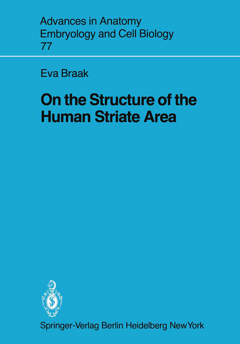
On the Structure of the Human Striate Area
Springer Berlin (Verlag)
978-3-540-11512-0 (ISBN)
1 Introduction.- 2 Material and Methods.- 3 Lamination Pattern.- 4 Neurons and Neuropil of Layer I.- 4.1 Nissl-Stained and Methylene Blue-Azure II-Stained Sections.- 4.2 Golgi Preparations.- 4.3 Electron Microscopy.- 5 General Remarks Concerning Pyramidal and Nonpyramidal or Stellate Cells.- 5.1 Nissl Preparations.- 5.2 Golgi Preparations.- 5.3 Electron Microscopy.- 6 Pyramidal Cells and Neuropil of Layer II.- 6.1 Nissl Preparations.- 6.2 Golgi Preparations.- 6.3 Electron Microscopy.- 7 Pyramidal Cells and Neuropil of Layer IIIab.- 7.1 Nissl Preparations.- 7.2 Golgi Preparations.- 7.3 Electron Microscopy.- 8 Pyramidal and Polygonal Neurons and Neuropil of Layers IIIc/IVa, IVb, IVc?, and IVc?.- 8.1 General Remarks.- 8.2 Nissl Preparations.- 8.3 Golgi Preparations.- 8.4 Electron Microscopy.- 8.5 Are the Polygonal Neurons Modified Pyramidal Neurons?.- 9 Pyramidal and Polygonal Neurons and Neuropil of Layers IVd/Va and Vb.- 9.1 Nissl-Stained and Methylene Blue-Azure II-Stained Sections.- 9.2 Golgi Preparations.- 9.3 Electron Microscopy.- 10 Pyramidal Cells and Multiformed Neurons of Layers VIa and VIb.- 10.1 Nissl Preparations.- 10.2 Golgi Preparations.- 10.3 Electron Microscopy.- 11 Nonpyramidal Cells of Layers II to VI.- 11.1 Golgi Preparations.- 11.2 Nissl Preparations.- 11.3 Electron Microscopy.- 12 Glial Cells of Layers I to VI.- 12.1 Astrocytes.- 12.2 Oligodendrocytes.- 12.3 Microglial Cells.- 13 Summary.- Acknowledgments.- References.
| Erscheint lt. Verlag | 1.7.1982 |
|---|---|
| Reihe/Serie | Advances in Anatomy, Embryology and Cell Biology |
| Zusatzinfo | VI, 87 p. 38 illus. |
| Verlagsort | Berlin |
| Sprache | englisch |
| Maße | 170 x 244 mm |
| Gewicht | 210 g |
| Themenwelt | Studium ► 1. Studienabschnitt (Vorklinik) ► Anatomie / Neuroanatomie |
| Schlagworte | Area striata • brain • Cell • electron microscopy • Ethylen • Microscopy • Structure |
| ISBN-10 | 3-540-11512-9 / 3540115129 |
| ISBN-13 | 978-3-540-11512-0 / 9783540115120 |
| Zustand | Neuware |
| Informationen gemäß Produktsicherheitsverordnung (GPSR) | |
| Haben Sie eine Frage zum Produkt? |
aus dem Bereich


