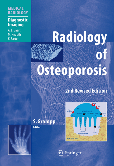
Radiology of Osteoporosis
Springer Berlin (Verlag)
978-3-642-06526-2 (ISBN)
This second edition of "Radiology of Osteoporosis" has been fully updated so as to represent the current state of the art. It provides a comprehensive overview of osteoporosis, the pathologic conditions that give rise to osteoporosis, and the complications that are frequently encountered. After initial chapters devoted to pathophysiology, the presentation of osteoporosis on conventional radiographs is illustrated and discussed. Thereafter, detailed consideration is given to each of the measurement methods employed to evaluate osteoporosis, including dual x-ray absorptiometry, vertebral morphometry, spinal and peripheral quantitative computed tomography, quantitative ultrasound, and magnetic resonance imaging. The role of densitometry in daily clinical practice is appraised. Finally, a collection of difficult cases involving pitfalls is presented, with guidance to their solution. The information contained in this volume will be invaluable to all with an interest in osteoporosis.
The revised second edition of Radiology of Osteoporosis has been fully updated to incorporate and analyze the current state of the art. It provides a comprehensive overview of osteoporosis, the pathologic conditions that give rise to osteoporosis, and frequently-encountered complications. Initial chapters are devoted to an overview of pathophysiology, followed by a thorough discussion and illustration of the presentation of osteoporosis on conventional radiographs. Detailed consideration is given to each of the measurement methods employed to evaluate osteoporosis, including dual x-ray absorptiometry, vertebral morphometry, spinal and peripheral quantitative computed tomography, quantitative ultrasound, and magnetic resonance imaging. The role of densitometry in daily clinical practice is appraised. Finally, the book presents a collection of difficult cases, with particular attention to their pitfals, and expert guidance on avoiding misinterpretation. This volume will be invaluable to all with an interest in osteoporosis.
to Bone Development, Remodelling and Repair.- Pathophysiology and Aging of Bone.- Pathophysiology of Rheumatoid Arthritis and Other Disorders.- Therapeutic Approaches and Mechanisms of Drug Action.- Orthopedic Surgery.- Radiology of Osteoporosis.- Dual-Energy X-Ray Absorptiometry.- Vertebral Morphometry.- Spinal Quantitative Computed Tomography.- pQCT: Peripheral Quantitative Computed Tomography.- Quantitative Ultrasound.- Magnetic Resonance Imaging.- Structure Analysis Using High-Resolution Imaging Techniques.- Densitometry in Clinical Practice.- Practical Cases.
From the reviews of the second edition:
"'Radiology of Osteoporosis,' ... is a revised edition of a volume in the Springer Medical Radiology series first published in 2002. The book is intended to provide a guide for radiologists, orthopaedic surgeons and other specialists on osteoporosis and the various medical imaging techniques that assist in its diagnosis. ... This book gives a useful and reasonably up-to-date account of the basic measurement techniques used in bone densitometry." (Glen Blake, RAD magazine, January, 2009)
"It provides a comprehensive overview of the different aspects of osteoporosis including the pathologic conditions that give rise to osteoporosis and the complications that frequently occur. Well-known international authors provide their most current data on morphology, pathophysiology and therapeutic approaches in osteoporosis ... . In summary, the information provided in this book will be invaluable to radiologists and all clinicians involved in the care of patients with osteoporosis." (A. K. Dixon and Thomas J. Vogl, European Radiology, Vol. 19, 2009)
| Erscheint lt. Verlag | 12.2.2010 |
|---|---|
| Reihe/Serie | Diagnostic Imaging | Medical Radiology |
| Vorwort | A.L. Baert |
| Zusatzinfo | X, 247 p. |
| Verlagsort | Berlin |
| Sprache | englisch |
| Maße | 193 x 270 mm |
| Gewicht | 570 g |
| Themenwelt | Medizinische Fachgebiete ► Radiologie / Bildgebende Verfahren ► Radiologie |
| Schlagworte | Computed tomography • Computed tomography (CT) • Densitometrie • densitometry • Fractures • Fraktura • Imaging • Imaging techniques • Magnetic Resonance • Magnetic Resonance Imaging • Magnetic Resonance Imaging (MRI) • Osteoporosis • pathophysiology • Physiology • quantitative computed tomography • quantitative CT • Radiology • Surgery • Tomography • Ultrasound • X-Ray |
| ISBN-10 | 3-642-06526-0 / 3642065260 |
| ISBN-13 | 978-3-642-06526-2 / 9783642065262 |
| Zustand | Neuware |
| Haben Sie eine Frage zum Produkt? |
aus dem Bereich


