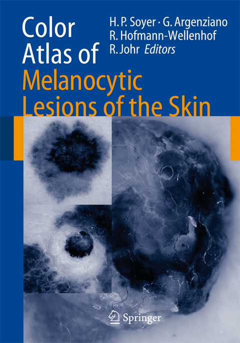
Color Atlas of Melanocytic Lesions of the Skin
Springer Berlin (Verlag)
978-3-642-07120-1 (ISBN)
Melanocytic tumors of the skin deserve special a ll these features characterize the book as an attention because of the following important impressive contribution to the literature in the facts area of melanocytic tumors. My co-workers in Graz, Dr. H. Peter Soyer ? Melanoma is frequent and early detection and Dr. r ainer Hofmann-Wellenhof, as well as is critical. Dr. Giuseppe a rgenziano from Naples and Dr. ? a correct interpretation is necessary r obert Johr from Miami, together with many because the implications may be very international contributors who are all experts in serious. their respective disciplines, have produced a ? it is a dynamically developing field where splendid piece of work which presents highly major progress has been made over the relevant information on a complex and cha- past decade. lenging subject. This book will greatly assist physicians in providing optimal care for pa - This atlas, written in a concise way, is a highly tients with melanocytic skin lesions.
Robert H. Johr, MD, Clinical Professor of Dermatology, Associate Clinical Professor of Pediatrics Pigmented Lesion Clinic, University of Miami School of Medicine, Miami, FL
The Morphologic Dimension in the Diagnosis of Melanocytic Skin Lesions.- Clinical Examination of Melanocytic Neoplasms Including ABCDE Criteria.- Dermoscopic Examination.- Melanoma: the Morphological Dimension.- Laser-Scanning Confocal Microscopy.- Automatic Diagnosis.- Multispectral Image Analysis.- Teledermatology.- The Life of Melanocytic Nevi.- Acral Nevus.- Agminated Nevus.- Blue Nevus.- Atypical (Dysplastic) Nevus.- Combined Nevus.- Common Nevus.- Congenital Melanocytic Nevi.- Melanocytic Nevi on the Genitalia and Melanocytic Nevi on Other Special Locations.- Halo Nevus.- Irritated Nevus and Meyerson's Nevus.- Melanocytic Lesions in Darker Racial Ethnic Groups.- Miescher Nevus.- Nevi with Particular Pigmentation: Black, Pink, and White Nevus.- Recurrent Nevus.- Spitz Nevus and Its Variants.- Syndromes Involving Melanocytic Lesions.- Nail Apparatus Nevus (Subungual Nevus, Nail Matrix Nevus).- Unna Nevus.- Epidemiology of Melanoma.- Acral Melanoma.- Amelanotic Melanoma.- Early Evolution of Melanoma (Small-Diameter Melanoma).- False-Negative Melanomas.- Genital Melanoma.- Melanoma of the Face.- Melanoma of the Trunk and Limbs Including Superficial and Nodular Melanoma.- Cutaneous Metastatic Melanoma.- Scalp Melanoma.- Nail Apparatus Melanoma (Subungual Melanoma, Nail Matrix Melanoma).- Pigmented Basal Cell Carcinoma.- Dermatofibroma.- Lentigines Including Lentigo Simplex, Reticulated Lentigo and Actinic Lentigo.- Squamous Cell Carcinoma Including Actinic Keratosis, Bowen's Disease, Keratoacanthoma, and Its Pigmented Variants.- Vascular Lesions.- Seborrheic Keratosis Including Lichen Planus-like Keratosis.
From the reviews:
"The purpose is to teach dermatologists how to increase diagnostic accuracy of pigmented lesions, especially melanoma, by using the dermatoscope. ... The audience is dermatologists and dermatology residents. ... This is definitely a valuable reference at a reasonable price. I would recommend reading it from cover to cover." (Patricia Wong, Doody's Review Service, March, 2009)
| Erscheint lt. Verlag | 14.10.2010 |
|---|---|
| Zusatzinfo | XVIII, 333 p. |
| Verlagsort | Berlin |
| Sprache | englisch |
| Maße | 170 x 242 mm |
| Gewicht | 595 g |
| Themenwelt | Medizin / Pharmazie ► Medizinische Fachgebiete ► Dermatologie |
| Schlagworte | carcinoma • Cell • Dermatology • Dermatopathology • Dermoscopy • Diagnosis • Differential Diagnosis • epidemiology • Laser • Laser-scanning in-vivo microscopy • Management • melanoma • Morphology • Pathology • Pigmented skin lesions • Skin |
| ISBN-10 | 3-642-07120-1 / 3642071201 |
| ISBN-13 | 978-3-642-07120-1 / 9783642071201 |
| Zustand | Neuware |
| Haben Sie eine Frage zum Produkt? |
aus dem Bereich


