Case-Based Nuclear Medicine
Thieme Medical Publishers Inc (Verlag)
978-1-58890-652-6 (ISBN)
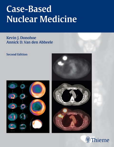
A highly visual clinical case review of nuclear medicine
Ideal for self-assessment, the second edition of Case-Based Nuclear Medicine has been fully updated to reflect the latest nuclear imaging technology, including cutting-edge cardiac imaging systems and the latest on PET/CT.
Each chapter is packed with high-quality images that demonstrate the full-range of commonly encountered disease manifestations as seen in the practice of nuclear medicine. The lavishly illustrated cases begin with the clinical presentation and a concise patient history followed by imaging findings, differential diagnoses, the definitive diagnosis and follow-up information, a brief discussion of the background for each diagnosis, and a list of pearls and pitfalls.
Features:
Comprehensive coverage of everything from single photon emission computed tomography to PET/CT imaging
Cases presented as 'unknowns' enable readers to develop their own differential diagnoses - just like on the exam
Over 400 high-resolution images, including full-color PET/CT and cardiac scintigraphic images, document the cases
Numerous tips, tricks, pearls, and pitfalls highlight key points at the end of each chapter
A scratch-off code provides 12 months of access to RadCases, a searchable online database of 250 must-know nuclear medicine cases
This user-friendly atlas is an essential resource for all residents and fellows in radiology and nuclear medicine as they review for exams and prepare for rounds. Clinicians will find the succinct presentation of cases an invaluable quick reference in daily practice.
Chief of the Department of Imaging, Founding Director of the Center of Biomedical Imaging in Oncology, and Director of Nuclear Medicine at the Dana-Farber Cancer Institute; Associate Professor, Department of Radiology, Harvard Medical School, Boston, Massachusetts, USA
Section I Skeletal Scintigraphy
Case 1 Normal Bone Scan
Case 2 Charcot Arthropathy
Case 3 Chondrosarcoma
Case 4 Ewing Sarcoma
Case 5 Metastatic Calcification
Case 6 Myocardial Uptake
Case 7 Paget Disease
Case 8 Renal Transplant
Case 9 Sacroiliitis
Case 10 Sickle Cell Disease
Case 11 Soft Tissue Metastasis
Case 12 Stress Fracture and Complex Regional Pain Syndrome
Case 13 Prostate Cancer
Case 14 Breast Cancer
Case 15 Lung Cancer
Case 16 Renal Cancer
Case 17 Superscan (Prostate Cancer)
Case 18 Liver Metastasis (Colon Cancer)
Case 19 Multiple Diseases
Case 20 Leiomyosarcoma
Case 21 Cold Defect (Lung Cancer)
Case 22 Ascites (Hepatoma)
Case 23 Radiation Therapy Port (Breast Cancer)
Case 24 Injection Infiltration
Case 25 Rib Trauma
Case 26 Paget Disease in Lumbar Vertebra
Case 27 Brain Abscess
Case 28 Stress Reaction (Shin Splints)
Case 29 Honda Sign
Case 30 Hip Prosthesis
Case 31 Pars Interarticularis Defect
Section II Cardiac Scintigraphy
Case 32 Normal SPECT
Case 33 Patient Motion on SPECT
Case 34 Breast Attenuation Artifact
Case 35 Diaphragmatic Attenuation Artifact
Case 36 Attenuation Artifact Corrected by Attenuation Correction
Case 37 Abnormal-Low Risk SPECT Scan
Case 38 Abnormal-High Risk SPECT Scan
Case 39 Normal PET
Case 40 Misregistration on PET
Case 41 Abnormal PET Scan
Case 42 FDG PET for Viability: Matched Defect
Case 43 FDG PET for Viability: Mismatched Defect
Section III Pulmonary Scintigraphy
Case 44 Normal Scintigraphy
Case 45 Very Low Likelihood
Case 46 Low Likelihood
Case 47 Intermediate Likelihood
Case 48 Intermediate Likelihood
Case 49 High Likelihood
Case 50 Intermediate Likelihood–X-Ray Match
Case 51 High Likelihood for Massive Pulmonary Embolism
Case 52 MAA Clumping
Case 53 Attenuation Artifact
Case 54 Right to Left Shunt–Atrial Septal Defect
Case 55 Right to Left Shunt–Pulmonary Hypertension
Section IV Endocrine Scintigraphy
Case 56 Normal Thyroid Gland
Case 57 Graves Disease and Cold Nodule
Case 58 Substernal Goiter
Case 59 Autonomous Adenoma
Case 60 Post-Partum Thyroiditis
Case 61 Whole-Body Survey Normal
Case 62 Pulmonary Metastasis
Case 63 Thymus
Case 64 Parathyroid Adenoma
Case 65 Mediastinal Parathyroid Adenoma
Case 66 Normal Parathyroid Bed
Case 67 Fifth Parathyroid Gland
Section V Scinitgraphy of Neoplastic Disease
Part A PET/CT
Case 68 Normal
Case 69 Lymphoma 1
Case 70 Lymphoma 2
Case 71 Lymphoma 3
Case 72 Lung Cancer 1
Case 73 Lung Cancer 2
Case 74 Lung Cancer 3
Case 75 Esophageal Cancer
Case 76 Colorectal Cancer 1
Case 77 Colorectal Cancer 2
Case 78 Pancreatic Cancer
Case 79 Breast Cancer 1
Case 80 Breast Cancer 2
Case 81 Breast Cancer 3
Case 82 Head and Neck Cancer 1
Case 83 Head and Neck Cancer 2
Case 84 Head and Neck Cancer 3
Case 85 Brain Cancer
Case 86 Thyroid Cancer
Case 87 Melanoma
Case 88 Ovarian Cancer
Case 89 Sarcoma
Case 90 Multiple Myeloma
Part B Neuroendocrine Imaging
Case 91 Carcinoid
Case 92 Pheochromocytoma
Case 93 Gastrinoma
Case 94 Neuroblastoma
Case 95 Insulinoma
Case 96 Multiple Endocrine Neoplasia
Case 97 Esthesioneuroblastoma
Section VI Radioisotope Therapy
Case 98 Thyroid Cancer 1
Case 99 Thyroid Cancer 2
Case 100 Hyperthyroidism
Case 101 Lymphoma
Case 102 Painful Bone Metastases
Case 103 Radiosynovectomy
Case 104 MIBG Therapy
Section VII Inflammation/Infection Imaging
Case 105 Normal
Case 106 Active Sarcoidosis
Case 107 Unilateral Sacroiliitis (with Osteomyelitis) and Adjacent Cellulitis
Case 108 Active Inflammatory Lesion without Organism Isolated
Case 109 Multifocal Infection
Case 110 Tibial Osteomyelitis
Case 111 Infected Pseudoaneurysm of Right Iliac Artery Endograft
Case 112 Peritonitis Related to Infected Peritoneal Dialysis Catheter
Section VIII Renal Scintigraphy
Case 113 Urine Leak
Case 114 Cortical Scintigraphy with DMSA
Case 115 Urinary Tract Obstruction
Case 116 Acute Tubular Necrosis
Case 117 GFR Calculation
Case 118 Glomerulonephritis
Case 119 Captopril Renography for Renovascular Hypertension
Section IX Biliary Scintigraphy
Case 120 Normal Hepatobiliary Scan
Case 121 Acute Gangrenous Cholecystitis
Case 122 Common Bile Duct Obstruction
Case 123 Gastric Bile Reflux
Case 124 Viral Hepatitis
Case 125 Bile Leak
Case 126 Space Occupying Masses in the Liver
Case 127 Gallbladder Ejection Fraction
Section X Lymphoscintigraphy
Case 128 Upper Extremity
Case 129 Lower Extremity
Case 130 Melanoma 1
Case 131 Melanoma 2
Section XI CNS Scintigraphy
Case 132 Alzheimer Disease
Case 133 Seizure
Case 134 Recurrent Astrocytoma
Case 135 Brain Death
Case 136 Normal Brain Flow Study
Case 137 Isolated Mid-Brain Tracer Uptake
Case 138 Tumor Recurrence
Section XII Gastrointestinal Scintigraphy
Case 139 Achalasia
Case 140 Reflux
Case 141 Normal Solid Gastric Emptying
Case 142 Gastroparesis
Case 143 Rapid Gastric Emptying
Case 144 Liquid Gastric Emptying
Case 145 Colonic Transit
Case 146 Meckel's Diverticulum
Case 147 Cecal Bleed/Colon Carcinoma
Case 148 Distal Colon Bleed/Colon Diverticulum
Case 149 Small Bowel Bleed/Duodenal Ulcer
Section XIII Vascular Scintigraphy
Case 150 Hemangioma 1
Case 151 Hemangioma 2
Case 152 Splenule 1
Case 153 Splenule 2
Case 154 Meckel 1
Case 155 Meckel 2
Case 156 Flow 1
Case 157 Flow 2
Case 158 Flow 3
Section XIV Pediatric Scintigraphy
Case 159 Urinary Tract Infection
Case 160 Meckel Diverticulum
Case 161 Biliary Atresia
Case 162 Epilepsy
Case 163 Neuroblastoma
Case 164 Acute Osteomyelitis
Case 165 Osteosarcoma
Case 166 Urinary Tract Obstruction
Appendix
Properties of Radioisotopes
| Erscheint lt. Verlag | 15.7.2011 |
|---|---|
| Zusatzinfo | 442 Illustrations |
| Verlagsort | New York |
| Sprache | englisch |
| Maße | 216 x 279 mm |
| Gewicht | 1814 g |
| Themenwelt | Medizinische Fachgebiete ► Radiologie / Bildgebende Verfahren ► Nuklearmedizin |
| Schlagworte | Atlas • Lehrbuch • Nuklearmedizin • Weiterbildung |
| ISBN-10 | 1-58890-652-3 / 1588906523 |
| ISBN-13 | 978-1-58890-652-6 / 9781588906526 |
| Zustand | Neuware |
| Haben Sie eine Frage zum Produkt? |
aus dem Bereich


