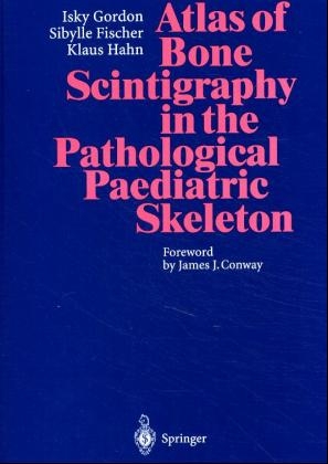
Atlas of Bone Scintigraphy in the Pathological Paediatric Skeleton
Springer Berlin (Verlag)
978-3-540-60471-6 (ISBN)
- Titel ist leider vergriffen;
keine Neuauflage - Artikel merken
This atlas covers both the common and the less common pathologies affecting the paediatric skeleton, providing illustrations, teaching points, technical comments, and text discussion. Variations in the appearances of osteomyelitis are also extensively illustrated. The procedures employed to create the images presented in this volume include whole body scanning, gamma camera high resolution spot images, pin hole and SPECT. Three phase bone scans are also illustrated. Indications for the use of each procedure are dicussed. The many illustrations in this atlas offer the paediatrician, orthopaedic surgeon, radiologist and nuclear medicine physician the opportunity to compare them with their own images or with the "normal" images presented in the previous companion volume.
Sibylle Fischer, BA Pädagogik der Frühen Kindheit, Fachwirtin für Kitas, gibt Fortbildungen für Erzieherinnen und arbeitet als wissenschaftliche Mitarbeiterin an der Evangelischen Hochschule Freiburg.
1 Introduction 2 Infection 2.1 Typical Hot Lesion in Bone 2.2 Less Common Appearances 2.3 Unusual Sites, Excluding the Long Bones 2.4 Non-skeletal Infection 2.5 Growth Arrest 3 Arthritis 3.1 Septic Arthritis 3.2 Aseptic Arthritis 4 Tumours 4.1 Benign Tumours 4.2 Malignant Tumours 4.3 Tumour Secondaries 4.4 Langerhans' Histiocytosis 5 Trauma 5.1 Appearances at Common Sites 5.2 Unusual Appearances of Locations of Fractures 5.3 Diffuse Skeletal Involvement 5.4 Complication of Trauma 5.5 Bone Response to Underlying Pathology 5.6 Post-operative Appearances 5.7 Effect of Radiotherapy 6 Osteochondritis Dissecans - Avascular Necrosis 6.1 Legg-Perthes' Disease 6.2 Other Sites of Osteochondritis Dissecans 6.3 Sickle Cell Disease 7 Miscellaneous 7.1 Dysplasia 7.2 Chondromata 7.3 Blount's Disease 7.4 Gaucher's Disease 7.5 Scoliosis 7.6 The Sick Child 7.7 Disuse Arthropathy 7.8 Muscular Disorders 7.9 Growth Arrest 8 Unusual Appearances of the Bone-Seeking Tracer 8.1 Kidney and Collecting System 8.2 Lung Uptake 8.3 Splenic Uptake 8.4 Brain Uptake 8.5 Soft Tissue Calcification 8.6 Isotope Artefact - Subject Index
| Vorwort | J. J. Conway |
|---|---|
| Sprache | englisch |
| Maße | 193 x 270 mm |
| Gewicht | 1196 g |
| Einbandart | gebunden |
| Themenwelt | Medizin / Pharmazie ► Medizinische Fachgebiete |
| Schlagworte | HC/Medizin/Klinische Fächer • Kinderheilkunde • Kinderheilkunde; Orthopädie • Knochenszintigraphie • Skelett |
| ISBN-10 | 3-540-60471-5 / 3540604715 |
| ISBN-13 | 978-3-540-60471-6 / 9783540604716 |
| Zustand | Neuware |
| Haben Sie eine Frage zum Produkt? |
aus dem Bereich
