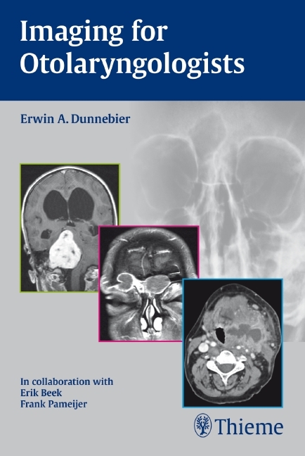
Imaging for Otolaryngologists
Seiten
2011
Thieme (Verlag)
978-3-13-146331-9 (ISBN)
Thieme (Verlag)
978-3-13-146331-9 (ISBN)
lt;br>A practical imaging primer designed specifically for ENTs
Imaging for Otolaryngologists distils the essentials of otolaryngologic imaging into a concise reference that concentrates on key topics that are of immediate interest to otolaryngologists practicing in a modern clinical environment.
Prepared by a renowned otolaryngologist, and reviewed and supplemented by expert radiologists, the book provides a well-rounded perspective. The central focus is on image interpretation, including the disease-specific characteristics, the features necessary for successful diagnosis, and the implications for surgery. Each of the 465 high-quality images is clearly labeled, and where appropriate comparisons are made between CT scans and MR images to show complementary functions and limitations.
Imaging for Otolaryngologists helps readers:
- Evaluate the cross-sectional anatomy in rhinology, otology, and laryngology on plain films, CT scans, and MR images- Appreciate the contribution and limitations of plain films, CT, and MRI in the management of otolaryngologic diseases- Select the best imaging modality for chronic, acute, and emergency otolaryngologic conditions- Understand which radiological appearances to look for in the diagnosis of common and less common otolaryngologic diseases
lt;p>General
1 Radiographic Imaging Techniques and Interpretation
Temporal Bone
2 Radiologic Anatomy of the Temporal Bone
3 Pathology of the Temporal Bone
Skull Base
4 Radiologic Anatomy of the Skull Base
5 Pathology of the Skull Base
Nose
6 Radiologic Anatomy of the Nasal Cavity and Paranasal Sinuses
7 Pathology of the Nasal Cavity and Paranasal Sinuses
Neck
8 Radiologic Anatomy of the Neck
9 Pathology of the Neck
| Erscheint lt. Verlag | 12.1.2011 |
|---|---|
| Verlagsort | Stuttgart |
| Sprache | englisch |
| Maße | 127 x 190 mm |
| Gewicht | 477 g |
| Themenwelt | Medizin / Pharmazie ► Medizinische Fachgebiete ► HNO-Heilkunde |
| Medizin / Pharmazie ► Medizinische Fachgebiete ► Radiologie / Bildgebende Verfahren | |
| Schlagworte | acute illnesses • akute Erkrankungen • Anatomie • anatomy • Bildgebende Verfahren • Bildinterpretationen • Chirurgie • choice of imaging procedure • chronic diseases • chronische Erkrankungen • common findings • Computed tomography • Computertomographie • COMPUTERTOMOGRA PHIE • CT • Diagnostic • Diagnostik • Differential Diagnosis • Differenzialdiagnose • disease findings • Diseases • Emergencies • ENT medicine • Erkrankungen • Hals • Hals-Nasen-Ohren-Heilkunde • Häufige Befunde • HÄUFIGE B EFUNDE • Head • HNO-Heilkunde • Image interpretations • imaging procedures • Injuries • Innenohr • Inner Ear • Kopf • Krankheitsbefunde • Magnetic Resonance Tomography • Magnetresonanztomogiraphie • MRT • nasal sinuses • Nasennebenhöhlen • neck • Normalanatomie • Normal anatomy • Notfälle • Rare findings • Röntgen • Röntgenanatomie • Schädelbasis • Schläfenbein • Seltene Befunde • Skull base • Surgery • temporary bone • Verletzungen • Wahl des bildgebenden Verfahrens • X-ray anatomy |
| ISBN-10 | 3-13-146331-7 / 3131463317 |
| ISBN-13 | 978-3-13-146331-9 / 9783131463319 |
| Zustand | Neuware |
| Haben Sie eine Frage zum Produkt? |
Mehr entdecken
aus dem Bereich
aus dem Bereich
ein Kompendium von Praktikern für Praktiker
Buch (2023)
Thieme (Verlag)
330,00 €
Differenzierte Diagnostik und Therapie
Buch | Hardcover (2021)
Springer (Verlag)
179,99 €
Buch | Softcover (2022)
Median-Verlag von Killisch-Horn GmbH
53,00 €


