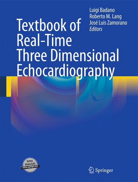
Textbook of Real-Time Three Dimensional Echocardiography
Springer London Ltd (Verlag)
978-1-84996-494-4 (ISBN)
- Titel erscheint in neuer Auflage
- Artikel merken
This Textbook will give the reader a detailed understanding of the use of 3D echo covering a wide range of topics; from the evolution of RT3D echo to the role of RT3D echo in drug trials, including chapters on the Principles of Transthoracic and Transesophageal Real-time 3D echocardiography. Other books in this area are more varied, less specific.
1. The evolution of three dimensional echocardiography.- Extrinsic tracking: positional and freehand acquisition.- Gated sequential imaging.- Integrated transesophageal approach and rotational transducer.- Real-time imaging.- 2. Principles of transthoracic three-dimensional acquisition and image display.- Matrix transducer design and technology.- Beam forming in three spatial dimensions.- Data processing .- Image resolution and display.- Image artifacts.- Three-dimensional color Doppler display and quantification.- Measuring in three dimensions.- Future developments.- 3. Principles of transesophageal three-dimensional acquisition and image display.- Matrix transducer design and technology.- Beam forming in three spatial dimensions.- Data processing.- Image resolution and display.- Image artifacts.- Unique quantification opportunities by transesophageal approach.- Future developments.- 4. Three-dimensional echocardiography in clinical practice.- Advantages over the two-dimensional modality.- Orientation of the heart and imaging views.- Acquisition modes and image display.- Implementing 3D echo in routine clinical practice.- 5. Three-dimensional echocardiographic evaluation of the left ventricle.- Validation studies.- Assessment of volume, shape and function.- Assessment of regional wall motion.- Assessment of mass.- Contrast enhancement.- Future directions.- 6. Advanced evaluation of left ventricular function with three dimensional echocardiography.- Assessment of intraventricular dyssynchrony.- Stress echocardiography.- Assessment of myocardial deformation .- Assessment of myocardial perfusion.- Future directions.- 7. Three-dimensional echocardiographic evaluation of the mitral valve.- Three dimensional echocardiographic anatomy of the normal mitral valve.- Mitral valve regurgitation.- Organic mitral valve regurgitation.- Functional mitral valve regurgitation.- Assessment of the degree of regurgitation.- Mitral valve stenosis.- Morphological assessment.- Functional assessment.- Monitoring percutaneous mitral valvuloplasty.- Mitral valve endocarditis.- 8.Three-dimensional echocardiographic evaluation of the aortic valve and the left ventricular outflow tract.- Three dimensional echocardiographic anatomy of the normal aortic valve.- The bicuspid aortic valve.- Assessment of aortic valve stenosis.- Assessment of aortic valve regurgitation.- Aortic valve endocarditis.- 9. Three dimensional echocardiographic evaluation of the right heart chambers.- The right ventricle.- Anatomy and function.- The tricuspid valve.- Normal anatomy.- Tricuspid regurgitation.- Tricuspid stenosis.- Tricuspid valve endocarditis.- The pulmonary valve.- Normal anatomy.- Pulmonary valve disease.- Pulmonary valve endocarditis.- 10. Three dimensional echocardiography in congenital heart disease.- 11. Three dimensional echocardiography to assess intracardiac masses.- Thrombi.- Benign Cardiac Tumors.- Malignant Cardiac Tumors.- Metastatic Tumors.- Devices.- 12. Use of three-dimensional echocardiography to guide intracardiac procedures.- 13. Future developments of three-dimensional echocardiography.- 14. Real-time three-dimensional transesophageal echocardiography.- 15. Real-time three dimensional echocardiography in mitral valve repair procedures.- 16. Visualization and assessment of coronary arteries with real-time three dimensional echocardiography.- 17. Three-dimensional echocardiography to assess cardiac geometry and function in athletes.- 18 Role of three-dimensional echo in drug trials.- Heart Failure.- Hypertension.
| Zusatzinfo | 36 Tables, black and white; 300 Illustrations, color; 100 Illustrations, black and white; XII, 198 p. 400 illus., 300 illus. in color. |
|---|---|
| Verlagsort | England |
| Sprache | englisch |
| Maße | 210 x 279 mm |
| Gewicht | 784 g |
| Themenwelt | Medizinische Fachgebiete ► Chirurgie ► Herz- / Thorax- / Gefäßchirurgie |
| Medizinische Fachgebiete ► Innere Medizin ► Kardiologie / Angiologie | |
| Medizinische Fachgebiete ► Radiologie / Bildgebende Verfahren ► Radiologie | |
| ISBN-10 | 1-84996-494-7 / 1849964947 |
| ISBN-13 | 978-1-84996-494-4 / 9781849964944 |
| Zustand | Neuware |
| Haben Sie eine Frage zum Produkt? |
aus dem Bereich
