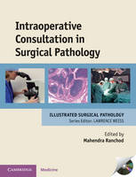
Intraoperative Consultation in Surgical Pathology
Cambridge University Press (Verlag)
978-0-521-89767-9 (ISBN)
Although frozen section diagnosis has been an integral part of surgical pathology for decades, this is the first textbook that offers a comprehensive clinicopathologic approach to the challenges of intraoperative consultation. Intraoperative diagnosis is challenging because of time constraints, sampling limitations, and the inability to perform a wide range of ancillary tests. Intraoperative Consultation in Surgical Pathology emphasizes the importance of clinical information, highlights the value of close collaboration with surgeons, and provides clear guidelines for the best way to examine specimens intraoperatively. Pathologists are then able to minimize error and diagnose with greater confidence. Most chapters in this book are co-authored by surgeons to ensure that their interests are represented. Essential reading for practising surgical pathologists, residents and fellows in pathology, this book will also be of value to fellows and surgeons in various surgical specialties who request intraoperative consultation.
Mahendra Ranchod is Adjunct Clinical Professor of Pathology, Stanford University Medical Center, Stanford and Director, Calpath and Gyne-Path Laboratories, Los Gatos, and Director of Anatomic Pathology, Good Samaritan Hospital, San Jose, California, USA.
Preface; 1. Introduction Mahendra Ranchod; 2. Technical aspects of intraoperative diagnosis Mahendra Ranchod; 3. Skin Mahendra Ranchod, Michael B. Morgan and Isaac M. Neuhaus; 4. Upper aerodigestive tract Raja R. Seethala, Mahendra Ranchod and Umamaheswar Duvvuri; 5. Lung Charles M. Lombard and Linda W. Martin; 6. Mediastinum Saul Suster, César A. Moran and David C. Rice; 7. Salivary glands Mahendra Ranchod, John K. C. Chan and David W. Eisele; 8. Gastrointestinal tract Steven D. Hart and Sarah M. Dry; 9. Liver, gallbladder and extrahepatic biliary tract Julia C. Iezzoni, Timothy M. Schmitt and Reid B. Adams; 10. Pancreas Ralph H. Hruban, Jon M. Davison, Richard D. Schulick, Karen M. Horton, Elliot K. Fishman and Syed Z. Ali; 11. Thyroid gland Mahendra Ranchod, John K. C. Chan and Electron Kebebew; 12. Parathyroid gland Mahendra Ranchod, John K. C. Chan and Electron Kebebew; 13. Breast Mahendra Ranchod, Roderick R. Turner and Carl A. Bertelsen; 14. Female genital tract Teri A. Longacre, Jonathan S. Berek and Michael R. Hendrickson; 15. Urinary tract and male genital system Steven S. Shen, Luan D. Truong, Seth P. Lerner and Jae Y. Ro; 16. Lymph nodes, spleen and extranodal lymphomas Patrick A. Treseler and Mahendra Ranchod; 17. Central nervous system Fausto J. Rodriguez, Bernd W. Scheithauer and John L. Atkinson; 18. Soft tissue Jesse K. McKenney, Raffi S. Avedian and Richard L. Kempson; 19. Bone and joint Andrew E. Horvai, Richard J. O'Donnell and Richard L. Kempson; 20. Pediatric surgical pathology Robert H. Byrd, Darrell L. Cass and Megan K. Dishop; Index.
| Erscheint lt. Verlag | 28.10.2010 |
|---|---|
| Reihe/Serie | Cambridge Illustrated Surgical Pathology |
| Zusatzinfo | 57 Tables, black and white; 344 Halftones, color; 3 Halftones, black and white; 21 Line drawings, color |
| Verlagsort | Cambridge |
| Sprache | englisch |
| Maße | 221 x 286 mm |
| Gewicht | 1500 g |
| Themenwelt | Medizin / Pharmazie ► Medizinische Fachgebiete ► Chirurgie |
| Studium ► 2. Studienabschnitt (Klinik) ► Pathologie | |
| ISBN-10 | 0-521-89767-X / 052189767X |
| ISBN-13 | 978-0-521-89767-9 / 9780521897679 |
| Zustand | Neuware |
| Haben Sie eine Frage zum Produkt? |
aus dem Bereich


