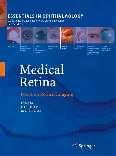
Medical Retina
Springer Berlin (Verlag)
978-3-540-85539-2 (ISBN)
Common Pitfalls in the Use of Optical Coherence Tomography for Macular Diseases.- Simultaneous SD-OCT and Confocal SLO-Imaging.- Fluorescein Angiography.- Wide-Field Imaging and Angiography.- Autofluorescence Imaging.- Imaging the Macular Pigment.- Near-Infrared Autofluorescence Imaging.- Near-Infrared Subretinal Imaging in Choroidal Neovascularization.- RetCam(TM) Imaging of Pediatric Intraocular Tumors.- Metabolic Mapping.- Assessing Diabetic Macular Edema with Optical Coherence Tomography.- OCT vs. Photography or Biomicroscopy for Diagnosis of Diabetic Macular Edema.- Spectral Domain Optical Coherence Tomography for Macular Holes.- Combined Spectral-Domain Optical Coherence Tomography/Scanning Laser Ophthalmoscopy Imaging of Vitreous and the Vitreo-Retinal Interface.- Choroidal Imaging with Optical Coherence Tomography.- Spectral-Domain Optical Coherence Tomography in Central Serous Chorioretinopathy.- New Developments in Optical Coherence Tomography Technology.- Toward Molecular Imaging.
From the reviews:
"This book does an outstanding job of describing established and new forms of retinal imaging, covering both acquisition and interpretation, information useful for clinicians as well as researchers. ... useful for those at all levels interested in applications of retinal imaging, from students and residents to experienced retina specialists. ... The figures and photographs illustrate important points well, and tables provide high-yield information in a easy to understand format. This would make an excellent addition to the library of anyone interested in retinal imaging." (Sonia Mehta, Doody's Review Service, September, 2010)
"The intended audience for Medical Retina consists of residents, clinicians, and basic science investigators. This particular volume of Medical Retina subtitled Focus on Retinal Imaging is testimony to the seminal advances made in retinal imaging; in particular scanning laser ophthalmoscopy (SLO) and optical low-coherence optical tomography (OCT). ... each chapter contains the key and up-to-date in-text citations to the end of chapter References which lists the complete citation including the first and the last pages. A detailed index completed the book." (Barry R. Masters, Graefe's Archive for Clinical and Experimental Ophthalmology, Vol. 249 (11), 2011)
"The book acts as a bridging text enabling the reader know of the recent advances and original research which can be used in day to day practice. ... text is laid out in a nice way. Each chapter starts with the core messages and gives a summary for clinicians. ... I would recommend this book not only for ophthalmologists involved in retina service ... but also for other ophthalmologists who would like to unravel the maze of the recent advances in the imaging of retina." (Atul Varma, Eye News, February/March, 2011)
| Erscheint lt. Verlag | 18.3.2010 |
|---|---|
| Reihe/Serie | Essentials in Ophthalmology |
| Zusatzinfo | XVI, 227 p. |
| Verlagsort | Berlin |
| Sprache | englisch |
| Maße | 193 x 260 mm |
| Gewicht | 688 g |
| Themenwelt | Medizin / Pharmazie ► Medizinische Fachgebiete ► Augenheilkunde |
| Medizinische Fachgebiete ► Radiologie / Bildgebende Verfahren ► Radiologie | |
| Schlagworte | AMD • Diagnosis • fluorescence • Laser • macular degeneration • Macular Disease • Molecular Imaging • Netzhaut • Optical Coherence Tomography (OCT) • retina • Retinal Imaging • Tomography • Tumor |
| ISBN-10 | 3-540-85539-4 / 3540855394 |
| ISBN-13 | 978-3-540-85539-2 / 9783540855392 |
| Zustand | Neuware |
| Haben Sie eine Frage zum Produkt? |
aus dem Bereich


