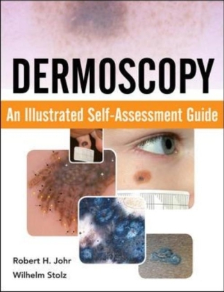
Dermoscopy: An Illustrated Self-Assessment Guide
Seiten
2010
McGraw-Hill Medical (Verlag)
978-0-07-161355-2 (ISBN)
McGraw-Hill Medical (Verlag)
978-0-07-161355-2 (ISBN)
- Titel erscheint in neuer Auflage
- Artikel merken
Zu diesem Artikel existiert eine Nachauflage
This unique resource helps readers train themselves in the use of direct skin microscopy (dermoscopy) in order to differentiate and diagnose pigmented skin lesions.
A case-based visual guide to learning dermoscopy complete with practical self-assessment
300 Full-color Illustrations —95 melanomas plus important simulators
A Doody's Core Title for 2011!
5 STAR DOODY'S REVIEW!
"This is a useful tool for any healthcare provider interested in dermatology, whether a new resident just starting to study skin disease or a seasoned practitioner wishing to learn a new skill....What is especially helpful is the labeling of the dermoscopic photo, making it easier to learn the various criteria and structures. Moreover, each case has a diagnostic pearls section that adds much didactic value....What is most unique and valuable is that readers are not just passively reading about a subject, but are drawn to actively participate and make decisions. When they turn the page, they can then contemplate the explanations and determine areas where they need improvement. The only thing that would top this book would be to have one of the authors alongside the reader at the bedside examining a patient. I wholeheartedly recommend this book for anyone interested in learning dermoscopy."--Doody's Review Service
With 300 full-color illustrations of 191 separate cases commonly encountered in general dermatologic practice, Dermoscopy: An Illustrated Guide offers a unique check-list methodology for learning how to use dermoscopy to diagnose benign and malignant pigmented and non-pigmented skin lesions.
For each of the 191 cases, you will find a series of high-quality full-color clinical and dermoscopic images, each with a short history. Every case is followed by five true-or-false questions along with three check boxes to test your knowledge acquisition and decision-making ability on “Risk, Diagnosis, and Disposition.” Turn the page and the answers to the questions are provided in a unique, memorable manner in which the dermoscopic images are presented again. Circles, stars, boxes, and arrows appear in the image pointing out the important dermoscopic criteria of each case.
Features
Cases are taken from lesions of the scalp, face, nose, lips, ears, trunk, extremities, palms, soles, nails and genitaliaThe global features and local criteria for each case are highlighted, which the authors have found to be a valuable teaching tool The concepts of clinico-dermoscopic correlation , dermoscopic-pathologic correlation and dermoscopic differential diagnosis are employed throughout the book Each case includes a discussion of all of its salient features in a quick-read outline style and ends with a series of dermoscopic and/or clinical pearls based on the authors’ years of experience treating skin cancers Key dermoscopic principles are re-emphasized throughout the book to enhance the reader’s understanding and assimilation of the teaching points
Dermoscopy: An Illustrated Self-Assessment Guide offers a simple, innovative, and highly-visual approach to learning the general principles, terminology, and specific criteria of dermoscopy to enhance your ability to utilize this powerful tool, which is both tissue sparing and potentially lifesaving.
Robert H. Johr, MD, Clinical Professor of Dermatology Associate Clinical Professor of Pediatrics Pigmented Lesion Clinic University of Miami School of Medicine Miami, FL Wilhelm Stolz, MD, Director, Clinic of Dermatology, Allergology, and Environmental Medicine Hospital Munchen Schwabing Professor of Dermatology, Faculty of Medicine Ludwig-Maximilians-Universitat, Munich, Germany
Preface
Acknowledgments
1. Dermoscopy from A to Z
2. Scalp, Face, Nose, and Ears
3. Trunk and Extremities
4. Palms, Soles, Nails
5. Genitalia
Index
| Erscheint lt. Verlag | 16.9.2010 |
|---|---|
| Verlagsort | New York |
| Sprache | englisch |
| Maße | 216 x 277 mm |
| Gewicht | 1225 g |
| Themenwelt | Medizin / Pharmazie ► Allgemeines / Lexika |
| Medizin / Pharmazie ► Medizinische Fachgebiete ► Dermatologie | |
| ISBN-10 | 0-07-161355-2 / 0071613552 |
| ISBN-13 | 978-0-07-161355-2 / 9780071613552 |
| Zustand | Neuware |
| Haben Sie eine Frage zum Produkt? |
Mehr entdecken
aus dem Bereich
aus dem Bereich
Differentialdiagnostik und Therapie bei Kindern und Jugendlichen
Buch (2022)
Thieme (Verlag)
165,00 €
Allergene - Diagnostik - Therapie
Buch (2022)
Thieme (Verlag)
200,00 €
richtig verschreiben – individuell therapieren
Buch (2023)
Thieme (Verlag)
71,00 €


