Radiology 101
The Basics and Fundamentals of Imaging
Seiten
2009
|
3rd Revised edition
Lippincott Williams and Wilkins (Verlag)
978-1-60547-225-6 (ISBN)
Lippincott Williams and Wilkins (Verlag)
978-1-60547-225-6 (ISBN)
- Titel erscheint in neuer Auflage
- Artikel merken
Zu diesem Artikel existiert eine Nachauflage
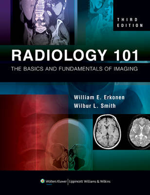
Featuring over 900 images, this book provides the basic groundwork necessary for interpreting images and understanding how imaging modalities function. It explains the principles, capabilities, and limitations of each imaging modality. It examines anatomic areas and organ systems, including a separate chapter on the pediatric chest and abdomen.
Featuring over 900 state-of-the-art images, "Radiology 101, Third Edition" provides the basic groundwork necessary for interpreting images and understanding how current imaging modalities function. The first chapter explains the principles, capabilities, and limitations of each imaging modality. Subsequent chapters examine anatomic areas and organ systems, including a separate chapter on the pediatric chest and abdomen. The books clearly labeled images show normal anatomy from various angles with various modalities and depict normal variants and common pathology. Each chapter includes suggested radiologic workups and key points summaries. This edition has extensive updates, especially on nuclear imaging (PET/CT), computed tomography (multi-slice), magnetic resonance (DWI, t-MRI, MRS), sonography (FAST), abdominal imaging (CT urography), mammography (digital mammograms), and the many indications for interventional radiology.
Featuring over 900 state-of-the-art images, "Radiology 101, Third Edition" provides the basic groundwork necessary for interpreting images and understanding how current imaging modalities function. The first chapter explains the principles, capabilities, and limitations of each imaging modality. Subsequent chapters examine anatomic areas and organ systems, including a separate chapter on the pediatric chest and abdomen. The books clearly labeled images show normal anatomy from various angles with various modalities and depict normal variants and common pathology. Each chapter includes suggested radiologic workups and key points summaries. This edition has extensive updates, especially on nuclear imaging (PET/CT), computed tomography (multi-slice), magnetic resonance (DWI, t-MRI, MRS), sonography (FAST), abdominal imaging (CT urography), mammography (digital mammograms), and the many indications for interventional radiology.
BASIC PRINCIPLES Radiography, Computed Tomography, Magnetic Resonance Imaging, Nuclear Imaging, and Ultrasonography: Basic Principles and Indications William E. Erkonen and Vincent A. Magnotta DIAGNOSTIC RADIOLOGY Chest William E. Erkonen and Brad H. Thompson Abdomen Edmund A. Franken, Jr. Pediatric Imaging Wilbur L. Smith Musculoskeletal System Carol A. Boles and William E. Erkonen Spine and Pelvis Carol A. Boles Brain Wilbur L. Smith Head and Neck Yutaka Sato Nuclear Imaging David L. Bushnell, Jr. Mammography William E. Erkonen, Laurie L. Fajardo, and Jeong Mi Park Interventional Radiology Thomas A. Farrell
| Mitarbeit |
Stellvertretende Herausgeber: Wilbur L. Smith |
|---|---|
| Zusatzinfo | Illustrations |
| Verlagsort | Philadelphia |
| Sprache | englisch |
| Maße | 213 x 277 mm |
| Gewicht | 1044 g |
| Themenwelt | Medizinische Fachgebiete ► Radiologie / Bildgebende Verfahren ► Radiologie |
| ISBN-10 | 1-60547-225-5 / 1605472255 |
| ISBN-13 | 978-1-60547-225-6 / 9781605472256 |
| Zustand | Neuware |
| Haben Sie eine Frage zum Produkt? |
Mehr entdecken
aus dem Bereich
aus dem Bereich
Nuklearmedizin und Strahlentherapie
Buch | Softcover (2024)
Springer (Verlag)
44,99 €


