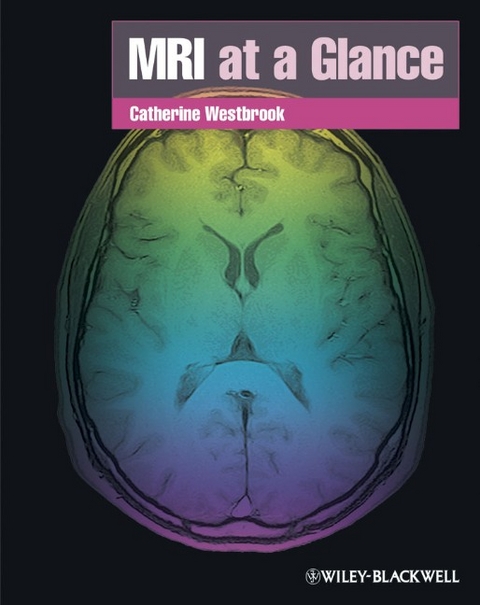
MRI at a Glance
Wiley-Blackwell (an imprint of John Wiley & Sons Ltd) (Verlag)
978-1-4051-9255-2 (ISBN)
- Titel erscheint in neuer Auflage
- Artikel merken
Catherine Westbrook MSc DCRR PgC (L&T) CTCert FHEA, is a senior lecturer and post-graduate pathway leader at the Faculty of Health & Social Care, Anglia Ruskin University Cambridge where she is responsible for post-graduate courses in MRI, CT, Radiography and Radiotherapy. Cathy is also an independent teaching consultant providing teaching and assessment in MRI and radiographic related subjects to clients all over the world.
Preface. Acknowledgements and Dedication. 1. Magnetism and electromagnetism. 2. Atomic structure. 3. Alignment and precession. 4. Resonance and signal generation. 5. Contrast mechanisms. 6. Relaxation mechanisms. 7. T1 recovery. 8. T2 decay. 9. T1 weighting. 10. T2 weighting. 11. Proton density weighting. 12. Pulse sequence mechanisms. 13. Conventional spin echo. 14. Fast or turbo spin echo - how it works. 15. Fast or turbo spin echo - how it's used. 16. Inversion recovery. 17. Gradient echo - how it works. 18. Gradient echo - how it's used. 19. The steady state. 20. Coherent gradient echo. 21. Incoherent gradient echo. 22. Steady-state free precession. 23. Balanced gradient echo. 24. Ultrafast sequences. 25. Diffusion and perfusion imaging. 26. Functional imaging techniques. 27. Gradient functions. 28. Slice selection. 29. Phase encoding. 30. Frequency encoding. 31. K space - what is it? 32. K space - how is it filled? 33. K space filling and signal amplitude. 34. K space filling and spatial resolution. 35. Data acquisition and frequency encoding. 36. Data acquisition and phase encoding. 37. Data acquisition and scan time. 38. K space traversal and pulse sequences. 39. Alternative K-space filling techniques. 40. Signal to noise ratio. 41. Contrast to noise ratio. 42. Spatial resolution. 43. Scan time. 44. Trade-offs. 45. Chemical shift. 46. Out-of-phase artefact. 47. Magnetic susceptibility. 48. Phase wrap/aliasing 94. 49. Phase mismapping (motion artefact). 50. Flow phenomena. 51. Time-of-flight MR angiography. 52. Phase contrast MR angiography. 53. Contrast enhanced MR angiography. 54. Contrast media. 55. Magnets. 56. Gradients. 57. Radiofrequency coils. 58. Other hardware. 59. Bioeffects. 60. Projectiles. 61. Screening and safety procedures. 62. Emergencies in the MR environment. Appendix 1. Appendix 2. Glossary. Index.
| Reihe/Serie | At a Glance |
|---|---|
| Zusatzinfo | Illustrations (mostly col.) |
| Verlagsort | Chicester |
| Sprache | englisch |
| Maße | 218 x 278 mm |
| Gewicht | 418 g |
| Themenwelt | Medizinische Fachgebiete ► Radiologie / Bildgebende Verfahren ► Kernspintomographie (MRT) |
| ISBN-10 | 1-4051-9255-0 / 1405192550 |
| ISBN-13 | 978-1-4051-9255-2 / 9781405192552 |
| Zustand | Neuware |
| Informationen gemäß Produktsicherheitsverordnung (GPSR) | |
| Haben Sie eine Frage zum Produkt? |
aus dem Bereich


