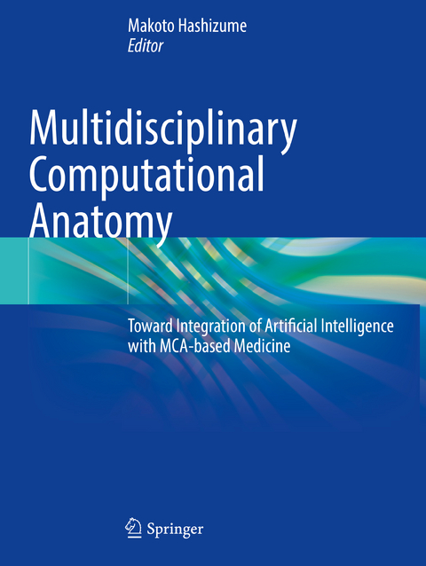
Multidisciplinary Computational Anatomy
Springer Verlag, Singapore
978-981-16-4327-9 (ISBN)
Multidisciplinary Computational Anatomy will appeal not only to clinicians but also to a wide readership in various scientific fields such as basic science, engineering, image processing, and biomedical engineering. All chapters were written byrespected specialists and feature abundant color illustrations. Moreover, the findings presented here share new insights into unresolved issues in the diagnosis and treatment of disease, and into the healthy human body.
Dr. Makoto Hashizume, MD, PhD, FACS, is a general surgeon who graduated at Kyushu University School of Medicine in 1979 and finished residency at General Surgery II, Kyushu University Hospital. Dr. Hashizume obtained his PhD in 1984 from Graduate School of Medical Sciences, Kyushu University. He was promoted to professor and chairman, Department of Disaster and Emergency Medicine, Faculty of Medical Sciences, Kyushu University, in 1999. He was awarded the title of distinguished professor in 2014 and the title of professor emeritus in 2018. Dr. Hashizume is now an executive director of Kitakyushu Koga Hospital in Japan.
Part I: Introduction: Perspectives toward MCA-based Medicine.- Ch 1: MCA: cancer detection, surgical and interventional sciences (David J Hawkes).- Part II: Basic Principles of MCA: Fundamental Theories and Techniques.- Ch 2: A Concept of Multi-disciplinary Computational Anatomy (MCA) (Yoshitaka Masutani).- Ch 3: Construction of Multi-Resolution Model of Pancreas Tumor (Hidekata Hontani).- Ch 4: Fundamental technologies for integration of multiscale spatiotemporal morphology in MCA (Akinobu Shimizu).- Ch 5: Fundamental technologies for integration and pathology in MCA (Yoshinobu Sato).- Part III: Basic Principles of MCA: Application Systems and Applied Techniques based on MCA Model.- Ch 6: Pre/intra-operative Diagnostic and Navigational Assistance Based on Multidisciplinary Computational Anatomy (Kensaku Mori).- Ch 7: Cancer diagnosis and prognosis assistance based on MCA (Noboru Niki).- Ch 8: Function integrated diagnostic assistance based on MCA models (Hiroshi Fujita).- Part IV: Basic Principles of MCA: Clinical and Scientific Application of MCA.- Ch 9: Clinical applications of MCA to surgery (Makoto Hashizum).- Ch 10: Clinical applications of MCA to diagnosis (Soji Kido).- Ch 11: Application of MCA across biomedical engineering (Etsuko Kobayashi).- Part V: New Frontier of Technology in Clinical Applications based on MCA Models: Lifelong Human Growth.- Ch 12: Three-dimensional analyses of human organogenesis (Tetsuya Takakuwa).- Ch 13: Skeletal system analysis during the human embryonic period based on MCA (Tetsuya Takakuwa).- Ch 14: MCA-based Embryology and Embryo Imaging (Shigeto Yamada).- Ch 15: Modeling of congenital heart malformations with a focus on topology (Ryou Haraguchi).- Part VI: New Frontier of Technology in Clinical Applications based on MCA Models: Tumor Growth.- Ch 16: A technique for measuring the 3D deformation of a multiphase structure to elucidate the mechanism of tumor invasion (Yasuyuki Morita).- Ch 17: Construction of classifier of tumore cell types of pancreas cancer based on pathological images using deep learning (Naoaki Ono).- Part VII: New Frontier of Technology in Clinical Applications based on MCA Models: Cranial Nervous System .- Ch 18: Multi-modal and multi-scale image registration for property analysis of brain tumor (Takashi Ohnishi).- Ch 19: Brain MRI image analysis technologies and its application to medical image analysis of Alzheimer’s diseases (Koichi Ito).- Ch 20: A computer aided support system for deep brain stimulation by multidisciplinary brain atlas database (Kenichi Morooka).- Ch 21: Integrating bio-metabolism and structural changes for the diagnosis of dementia (Yuichi Kimura).- Ch 22: Normalized Brain Datasets with Functional Information Predict the Glioma Surgery (Manabu Tamura).- Part VIII: New Frontier of Technology in Clinical Applications based on MCA Models: Cardio-respiratory System.- Ch 23: MCA analysis for the change in the cardiac fiber orientation under congestive heart failure(Toshiaki Akita).- Ch 24: Computerized evaluation of pulmonary function based on the rib and diaphragm motion by dynamic chest radiography (Rie Tanaka).- Ch 25: Computer-aided diagnosis of interstitial lung disease on high-resolution CT imaging parallel to the chest (Shingo Iwano).- Ch 26: Postoperative prediction of pulmonary resection based on MCA model by integrating the temporal responses and bio-mechanical functions (Fei Jiang).- Part IX: New Frontier of Technology in Clinical Applications based on MCA Models: Abdominal Organs.- Ch 27: Analysis in Three-dimensional Morphologies of Hepatic Microstructures in Hepatic Disease (Hiroto Shoji).- Ch 28: Quantitative Evaluation of Fatty Metamorphosis and Fibrosis of Liver Based on Models of Ultrasound and Light Propagation and its Application to Hepatic Disease Diagnosis (Tsuyoshi Shiina).- Ch 29: MCA analysis for hepatology~ Establishment of the in-situ visualization system for liver sinusoid analysis (Keiichi Akahoshi).- Ch 30: Simulation surgery for hepatobiliary-pancreatic surgery (Yukio Ohshiro).- Part X: New Frontier of Technology in Clinical Applications based on MCA Models: Musculoskeletal System.- Ch 31: Model Generation for Respiratory Muscle Function Analysis (Naoki Kamiya).- Ch 32: Morphometric analysis for the morphogenesis of the craniofacial structures and the evolution of the nasal protrusion in humans (Motoki Katsube).- Ch 33: Development of Bone Strength Prediction Method by Using MCA with Damage Mechanics (Mitsugu Todo).- Part XI: New Frontier of Technology in Clinical Applications based on MCA Models: Emerging Innovasive Imaging Technology.- Ch 34: Development of a generation method for local appearance models of normal organs by DCNN (Shouhei Hanaoka).- Ch 35: Modeling - Disorder development onset prediction based on spatiotemporal statistical shape model (Saadia Binte Alam).- Ch 36: Prediction of personalized post-operative implanted knee kin-ematics with statistical temporal modeling (Belayat Hossain).- Ch 37: Sparse Modeling in Analysis for Multidisciplinary Medical Data (Yen-Wei Chen).- Ch 38: Super Computing - Creation of large-scale and high-performance technology for processes of MCA by utilizing supercomputers (Takahiro Katagiri).- Ch 39: MRI - Quantitative evaluation of diseased tissue by viscoelastic imaging systems (Mikio Suga).- Ch 40: US - Development of general biophysical model for realization of ultrasonic qualitative real-time pathological diagnosis (Tadashi Yamaguchi).- Ch 41: OCT - Ultrahigh resolution optical coherence tomography at visible to near- infrared wavelength region (Norihiko Nishizawa).- Ch 42: MRI - Magnetic Resonance Q-Space Imaging Using Generating Function and Bayesian Inference (Eizou Umezawa).- Ch 43: Micro-CT and Lungs (Shota Nakamura).- Ch 44: Real-time endoscopic computer vision technologies and their applications that help improve the level of autonomy of surgi-cal assistant robots (Atsushi Nishikawa).- Ch 45: Endoscopy - Computer-aided diagnostic system based on deep learning which supports endoscopists’ decision making on the treatment of colorectal polyps (Yuichi Mori).- Ch 46: Endoscopy - Application of MCA modeling to abnormal nerve plexus in the GI tract (Kazuki Sumiyama).- Ch 47: Optical Fluorescence: Application of structured light illumination and compressed sensing to high speed laminar optical fluorescence tomography (Ichiro Sakuma).- Ch 48: Magneto-stimulation system for brain based on medical images (Akimasa Hirata).- Ch 49: AI - A machine-learning-based framework for developing various computer-aided detection systems with generated image features (Mitsutaka Nemoto).- Ch 50: Radiomics - Artificial intelligence based radiogenomic diagnosis of gliomas (Manabu Kinoshita).- Ch 51: 4D - Four-dimensional dynamic images: principle and future application (Naoki Suzuki).- Ch 52: Cloud XR (Extended Reality: Virtual Reality・Augmented Reality・Mixed Reality) and 5G networks for holographic medical image-guided surgeryand telemedicine (Maki Sugimoto).- Ch 53: Smart Cyber Operating Theater (SCOT)– Strategy for Future OR (Yoshihiro Muragaki).- Appendix: MCA models: Lifetime, Brain, Chest, Abdominal, Skeletomuscular system.
| Erscheinungsdatum | 07.12.2022 |
|---|---|
| Zusatzinfo | 228 Illustrations, color; 26 Illustrations, black and white; XV, 398 p. 254 illus., 228 illus. in color. |
| Verlagsort | Singapore |
| Sprache | englisch |
| Maße | 210 x 279 mm |
| Themenwelt | Mathematik / Informatik ► Informatik |
| Medizinische Fachgebiete ► Radiologie / Bildgebende Verfahren ► Radiologie | |
| Studium ► 1. Studienabschnitt (Vorklinik) ► Anatomie / Neuroanatomie | |
| Technik ► Medizintechnik | |
| Schlagworte | Artificial Intelligence • computational anatomy • computer-aided surgery • MCA • Medical Image Processing • robotic surgery • Surgical navigation system |
| ISBN-10 | 981-16-4327-X / 981164327X |
| ISBN-13 | 978-981-16-4327-9 / 9789811643279 |
| Zustand | Neuware |
| Haben Sie eine Frage zum Produkt? |
aus dem Bereich


