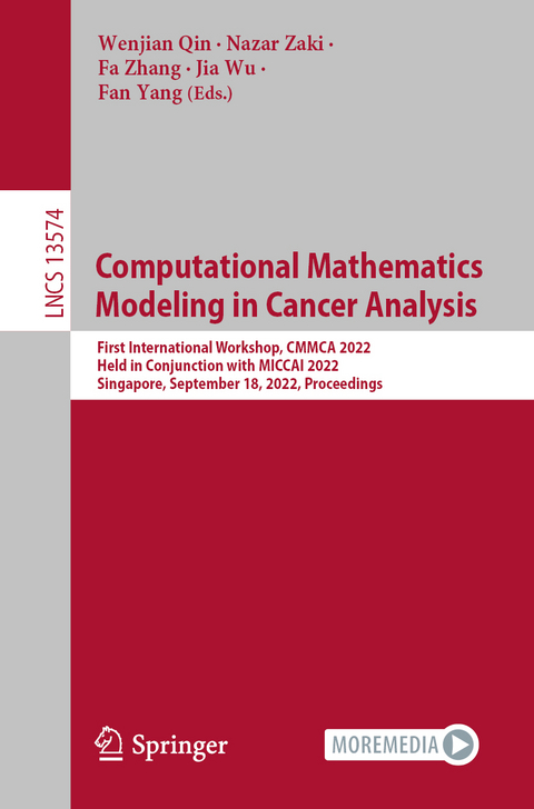
Computational Mathematics Modeling in Cancer Analysis
Springer International Publishing (Verlag)
978-3-031-17265-6 (ISBN)
This book constitutes the proceedings of the First Workshop on Computational Mathematics Modeling in Cancer Analysis (CMMCA2022), held in conjunction with MICCAI 2022, in Singapore in September 2022. Due to the COVID-19 pandemic restrictions, the CMMCA2022 was held virtually.
DALI 2022 accepted 15 papers from the 16 submissions that were reviewed. A major focus of CMMCA2022 is to identify new cutting-edge techniques and their applications in cancer data analysis in response to trends and challenges in theoretical, computational and applied aspects of mathematics in cancer data analysis.
Cellular Architecture on Whole Slide Images Allows the Prediction of Survival in Lung Adenocarcinoma .- Is More Always Better? Effects of Patch Sampling in Distinguishing Chronic Lymphocytic Leukemia from Transformation to Diffuse Large B-cell Lymphoma.- Repeatability of Radiomic Features against Simulated Scanning Position Stochasticity across Imaging Modalities and Cancer Subtypes: A Retrospective Multi-Institutional Study on Head-and-Neck Cases.- MLCN: Metric Learning Constrained Network for Whole Slide Image Classification with Bilinear Gated Attention Mechanism.- NucDETR: End-to-End Transformer for Nucleus Detection in Histopathology Images.- Self-supervised learning based on a pre-trained method for the subtype classification of spinal tumors.- CanDLE: Illuminating Biases in Transcriptomic Pan-Cancer Diagnosis.- Cross-Stream Interactions: Segmentation of Lung Adenocarcinoma Growth Patterns.- Modality-collaborative AI model Ensemble for Lung Cancer Early Diagnosis.- Clustering-based Multi-instance Learning Network for Whole Slide Image Classification.- Multi-task Learning-driven Volume and Slice Level Contrastive Learning for 3D Medical Image Classification.- Light Annotation Fine Segmentation: Histology Image Segmentation based on VGG Fusion with Global Normalisation CAM.- Tubular Structure-Aware Convolutional Neural Networks for Organ at Risks Segmentation in Cervical Cancer Radiotherapy.- Automatic Computer-aided Histopathologic Segmentation for Nasopharyngeal Carcinoma using Transformer Framework.- Accurate Breast Tumor Identification UsingComputational Ultrasound Image Features.
| Erscheinungsdatum | 21.09.2022 |
|---|---|
| Reihe/Serie | Lecture Notes in Computer Science |
| Zusatzinfo | X, 160 p. 59 illus., 56 illus. in color. |
| Verlagsort | Cham |
| Sprache | englisch |
| Maße | 155 x 235 mm |
| Gewicht | 272 g |
| Themenwelt | Informatik ► Grafik / Design ► Digitale Bildverarbeitung |
| Schlagworte | Applications • Artificial Intelligence • cancer diagnosis • color image processing • Computer Science • Computer systems • computer vision • conference proceedings • Deep learning • Image Analysis • image matching • Image Processing • Image Segmentation • Informatics • machine learning • Mathematics • Neural networks • pattern recognition • reference image • Research • Signal Processing |
| ISBN-10 | 3-031-17265-5 / 3031172655 |
| ISBN-13 | 978-3-031-17265-6 / 9783031172656 |
| Zustand | Neuware |
| Haben Sie eine Frage zum Produkt? |
aus dem Bereich


