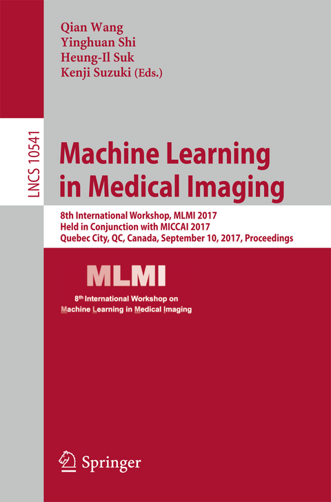
Machine Learning in Medical Imaging
Springer International Publishing (Verlag)
978-3-319-67388-2 (ISBN)
The 44 full papers presented in this volume were carefully reviewed and selected from 63 submissions. The main aim of this workshop is to help advance scientific research within the broad field of machine learning in medical imaging. The workshop focuses on major trends and challenges in this area, and presents works aimed to identify new cutting-edge techniques and their use in medical imaging.
From Large to Small Organ Segmentation in CT Using Regional Context.- Motion Corruption Detection in Breast DCE-MRI.- Detection and Localization of Drosophila Egg Chambers in Microscopy Images.- Growing a Random Forest with Fuzzy Spatial Features for Fully Automatic Artery-specific Coronary Calcium Scoring.- Atlas of Classifiers for Brain MRI Segmentation.- Dictionary Learning and Sparse Coding-based Denoising for High-Resolution Task Functional Connectivity MRI Analysis.- Yet Another ADNI Machine Learning Paper? Paving The Way Towards Fully-reproducible Research on Classification of Alzheimer's Disease.- Multi-Factorial Age Estimation from Skeletal and Dental MRI Volumes.- Automatic Classification of Proximal Femur Fractures Based on Attention Models.- Joint Supervoxel Classification Forest for Weakly-Supervised Organ Segmentation.- Accurate and Consistent Hippocampus Segmentation Through Convolutional LSTM and View Ensemble.- STAR: Spatio-Temporal Architecture for Super-Resolution inLow-Dose CT Perfusion.- Classification of Alzheimer's Disease by Cascaded Convolutional Neural Networks Using PET Images.- Finding Dense Supervoxel Correspondence of Cone-Beam Computed Tomography Images.- Multi-Scale Volumetric ConvNet with Nested Residual Connections for Segmentation of Anterior Cranial Base.- Feature Learning and Fusion of Multimodality Neuroimaging and Genetic Data for Multi-Status Dementia Diagnosis.- 3D Convolutional Neural Networks with Graph Refinement for Airway Segmentation Using Incomplete Data Labels.- Efficient Groupwise Registration for Brain MRI by Fast Initialization.- Sparse Multi-View Task-centralized Learning for ASD Diagnosis.- Inter-Subject Similarity Guided Brain Network Modelling for MCI Diagnosis.- Scalable and Fault Tolerant Platform for Distributed Learning on Private Medical Data.- Triple-Crossing 2.5D Convolutional Neural Network for Detecting Neuronal Arbours in 3D Microscopic Images.- Longitudinally-Consistent Parcellation of Infant Population Cortical Surfaces Based on Functional Connectivity.- Gradient Boosted Trees for Corrective Learning.- Self-paced Convolutional Neural Network for Computer Aided Detection in Medical Imaging Analysis.- A Point Says a Lot: An Interactive Segmentation Method for MR Prostate via One-Point Labeling.- Collage CNN for Renal Cell Carcinoma Detection from CT.- Aggregating Deep Convolutional Features for Melanoma Recognition in Dermoscopy Images.- Localizing Cardiac Structures in Fetal Heart Ultrasound Video.- Deformable Registration Through Learning of Context-Specific Metric Aggregation.- Segmentation of Craniomaxillofacial Bony Structures from MRI with a 3D Deep-learning Based Cascade Framework.- 3D U-net with Multi-Level Deep Supervision: Fully Automatic Segmentation of Proximal Femur in 3D MR Images.- Indecisive Trees for Classification and Prediction of Knee Osteoarthritis.- Whole Brain Segmentation and Labeling from CT using synthetic MR Images.- Structural Connectivity Guided SparseEffective Connectivity for MCI Identification.- Fusion of High-order and Low-order Effective Connectivity Networks for MCI Classification.- Novel Effective Connectivity Network Inference for MCI Identification.- Reconstruction of Thin-Slice Medical Images Using Generative Adversarial Network.- Neural Network Convolution (NNC) for Converting Ultra-Low-Dose to "Virtual" High-Dose CT Images.- Deep-Fext: Deep Feature Extraction for Vessel Segmentation and Centerline Prediction.- Product Space Decompositions for Continuous Representations of Brain Connectivity.- Identifying Autism from Resting-State fMRI Using Long Short-Term Memory Networks.- Machine Learning for Large-Scale Quality Control of 3D Shape Models in Neuroimaging.- Tversky Loss Function for Image Segmentation Using 3D Fully Convolutional Deep Networks.
| Erscheinungsdatum | 15.10.2017 |
|---|---|
| Reihe/Serie | Image Processing, Computer Vision, Pattern Recognition, and Graphics | Lecture Notes in Computer Science |
| Zusatzinfo | XV, 391 p. 134 illus. |
| Verlagsort | Cham |
| Sprache | englisch |
| Maße | 155 x 235 mm |
| Gewicht | 615 g |
| Themenwelt | Informatik ► Grafik / Design ► Digitale Bildverarbeitung |
| Schlagworte | Applications • classification and regression trees • Computer Science • computer vision • conference proceedings • convolutional neural networks • Deep learning • factorization methods • generative adversarial networks • Image Analysis • Image Processing • image processing and computer vision • Image Segmentation • Informatics • Learning Networks • Learning Systems • machine learning • Medical Image Analysis • multi-task learning • Neural networks • Research • Shape Representation • supervised learning |
| ISBN-10 | 3-319-67388-2 / 3319673882 |
| ISBN-13 | 978-3-319-67388-2 / 9783319673882 |
| Zustand | Neuware |
| Haben Sie eine Frage zum Produkt? |
aus dem Bereich


