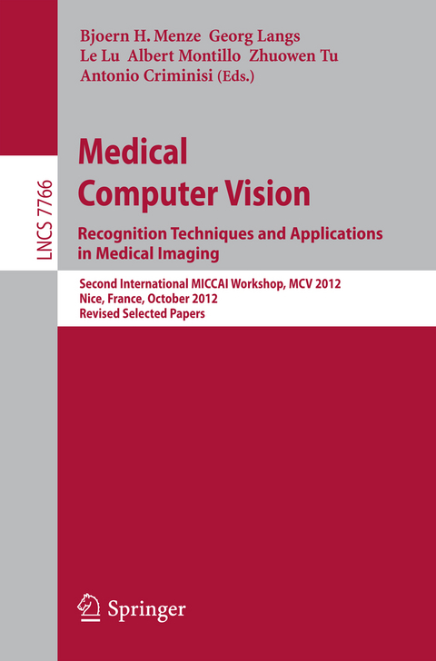
Medical Computer Vision: Recognition Techniques and Applications in Medical Imaging
Springer Berlin (Verlag)
978-3-642-36619-2 (ISBN)
The 24 papers have been selected out of 42 submissions. At MCV 2012, 12 papers were presented as a poster and 12 as a poster together with a plenary talk. The book also features four selected papers which were presented at the previous CVPR Medical Computer Vision workshop held in conjunction with the International Conference on Computer Vision and Pattern Recognition on June 21 2012 in Providence, Rhode Island, USA. The papers explore the use of modern computer vision technology in tasks such as automatic segmentation and registration, localization of anatomical features and detection of anomalies, as well as 3D reconstruction and biophysical model personalization.
Real-Time 2D/3D Deformable Registration Using Metric Learning.- Groupwise Spectral Log-Demons Framework for Atlas Construction.- Robust Anatomical Correspondence Detection by Graph Matching with Sparsity Constraint.- Semi-supervised Segmentation Using Multiple Segmentation Hypotheses from a Single Atlas.- Carotid Artery Wall Segmentation by Coupled Surface Graph Cuts.- Graph Cut Segmentation Using a Constrained Statistical Model with Non-linear and Sparse Shape Optimization.- Novel Context Rich LoCo and GloCo Features with Local and Global Shape Constraints for Segmentation of 3D Echocardiograms with Random Forests.- Novel Vector-Valued Approach to Automatic Brain Tissue Classification.- Atlas-Based Whole-Body PET-CT Segmentation Using a Passive Contour Distance.- Spatially Aware Patch-Based Segmentation (SAPS): An Alternative Patch-Based Segmentation Framework.- EfficientGeometrical Potential Force Computation for Deformable Model Segmentation.- Shape Prior Model for Media-Adventitia Border Segmentation in IVUS Using Graph Cut.- Multiple Atlases-Based Joint Labeling of Human Cortical Sulcal Curves.- Fast Anatomical Structure Localization Using Top-Down Image Patch Regression.- Oblique Random Forests for 3-D Vessel Detection Using Steerable Filters and Orthogonal Subspace Filtering.- Pipeline for Tracking Neural Progenitor Cells.- Automatic Heart Isolation in 3D CT Images.- Randomness and Sparsity Induced Codebook Learning with Application to Cancer Image Classification.- Context Enhanced Graphical Model for Object Localization in Medical Images.- A Cascade Learning Method for Liver Lesion Detection in CT Images.- Automatic Event Detection within Thrombus Formation Based on Integer Programming.- Automatic Extraction of the Curved Midsagittal Brain Surface on MR Images.- Identification of Malignant Breast Tumors Based on Acoustic Attenuation Mapping of Conventional Ultrasound Images.- What Genes Tell about Iris Appearance.-Robust Dense Endoscopic Stereo Reconstruction for Minimally Invasive Surgery.- Model-Based Human Teeth Shape Recovery from a Single Optical Image with Unknown Illumination.- Brain Tumor Cell Density Estimation from Multi-modal MR Images Based on a Synthetic Tumor Growth Model.- Current-Based 4D Shape Analysis for the Mechanical Personalization of Heart Models.
Groupwise Spectral Log-Demons Framework for Atlas Construction.- Robust Anatomical Correspondence Detection by Graph Matching with Sparsity Constraint.- Semi-supervised Segmentation Using Multiple Segmentation Hypotheses from a Single Atlas.- Carotid Artery Wall Segmentation by Coupled Surface Graph Cuts.- Graph Cut Segmentation Using a Constrained Statistical Model with Non-linear and Sparse Shape Optimization.- Novel Context Rich LoCo and GloCo Features with Local and Global Shape Constraints forSegmentation of 3D Echocardiograms with Random Forests.- Novel Vector-Valued Approach to Automatic Brain Tissue Classification.- Atlas-Based Whole-Body PET-CT Segmentation Using a Passive Contour Distance.- Spatially Aware Patch-Based Segmentation (SAPS): An Alternative Patch-Based Segmentation Framework.- Efficient Geometrical Potential Force Computation for Deformable Model Segmentation.- Shape Prior Model for Media-Adventitia Border Segmentation in IVUS Using Graph Cut.- Multiple Atlases-Based Joint Labeling of Human Cortical Sulcal Curves.- Fast Anatomical Structure Localization Using Top-Down Image Patch Regression.- Oblique Random Forests for 3-D Vessel Detection Using Steerable Filters and Orthogonal Subspace Filtering.- Pipeline for Tracking Neural Progenitor Cells.- Automatic Heart Isolation in 3D CT Images.- Randomness and Sparsity Induced Codebook Learning with Application to Cancer Image Classification.- Context Enhanced Graphical Model for Object Localization in Medical Images.- A Cascade Learning Method for Liver Lesion Detection in CT Images.-Automatic Event Detection within Thrombus Formation Based on Integer Programming.- Automatic Extraction of the Curved Midsagittal Brain Surface on MR Images.- Identification of Malignant Breast Tumors Based on Acoustic Attenuation Mapping of Conventional Ultrasound Images.- What Genes Tell about Iris Appearance.- Robust Dense Endoscopic Stereo Reconstruction for Minimally Invasive Surgery.- Model-Based Human Teeth Shape Recovery from a Single Optical Image with Unknown Illumination.- Brain Tumor Cell Density Estimation from Multi-modal MR Images Based on a Synthetic Tumor Growth Model.- Current-Based 4D Shape Analysis for the Mechanical Personalization of Heart Models.| Erscheint lt. Verlag | 27.3.2013 |
|---|---|
| Reihe/Serie | Image Processing, Computer Vision, Pattern Recognition, and Graphics | Lecture Notes in Computer Science |
| Zusatzinfo | XI, 294 p. 136 illus. |
| Verlagsort | Berlin |
| Sprache | englisch |
| Maße | 155 x 235 mm |
| Gewicht | 468 g |
| Themenwelt | Informatik ► Grafik / Design ► Digitale Bildverarbeitung |
| Informatik ► Theorie / Studium ► Künstliche Intelligenz / Robotik | |
| Schlagworte | atlas-based labeling • graph cut segmentation • machine learning • Medical Image Analysis • MR images |
| ISBN-10 | 3-642-36619-8 / 3642366198 |
| ISBN-13 | 978-3-642-36619-2 / 9783642366192 |
| Zustand | Neuware |
| Haben Sie eine Frage zum Produkt? |
aus dem Bereich
