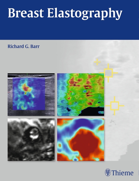
Breast Elastography
Thieme Medical Publishers Inc (Verlag)
978-1-60406-852-8 (ISBN)
This comprehensive reference covers the principles and techniques used in performing breast elastography, an innovative imaging technology that can dramatically reduce the need for biopsies. The book begins with an introduction of the techniques, followed by sections on how to perform each technique and methods of interpretation, and concludes with more than 60 detailed case studies.
Key Features:
Includes case studies covering a wide range of breast pathologies and illustrating the use of all available elastography techniques to help radiologists obtain the best images for each pathology
Covers all methods of breast elastography, including sheer wave and strain wave
Contains more than 200 high-quality color images that demonstrate how to perform each technique
Breast Elastography is an essential reference for all radiologists, residents and fellows, and sonographers involved in breast imaging and evaluation.
Introduction to Breast Elastography
2 Principles of Elastography
3 Strain Elastography
4 Shear Wave Elastography
5 Combination of Shear Wave and Strain Elastography
6 Clinical Cases: Benign Lesions
Case 1: Cystic Lesion—Simple Cyst
Case 2: Cystic Lesion—Complicated Cyst
Case 3: Cystic Lesion—Complicated Cyst
Case 4: Cystic Lesion Isoechoic Complicated Cyst
Case 5: Cystic Lesion—Apocrine Microcyst
Case 6: Cystic Lesion—Complex Cystic Lesion
Case 7: Cystic Lesion—Complex Cystic Lesion
Case 8: Fibrocystic Change—Lesion Blends in with Adjacent Tissue on Elastography
Case 9: Fibrocystic Change—Fibroadenomoid Hyperplasia
Case 10: Fibrocystic Change—Lesion Blends in with Adjacent Tissue on Elastography
Case 11: Fibrocystic Change—Stromal Fibrosis
Case 12: Sclerosing Lesion—Sclerosis Lobulitis
Case 13: Fibroadenoma—Soft Fibroadenoma
Case 14: Fibroadenoma—Intermediate Stiffness Fibroadenoma
Case 15: Fibroadenoma—Stiffer Fibroadenoma
Case 16: Fibroadenoma—Stiffer Fibroadenoma
Case 17: Fibroadenoma—False-Positive Fibroadenoma
Case 18: Papillary Lesion—Hyalinized Intraductal Papilloma
Case 19: Papillary Lesion—Intraductal Papilloma
Case 20: Papillary Lesion—Intraductal Papillomatosis
Case 21: Papillary Lesion—Intraductal Papilloma
Case 22: Phyllodes Tumor
Case 23: Mastitis—Mastitis with Abscess Formation
Case 24: Mastitis—Mastitis with Abscess Formation
Case 25: Mastitis—Mastitis without Abscess Formation
Case 26: Mastitis—Chronic Granulomatous Mastitis
Case 27: Surgical Scar—Benign Surgical Scar
Case 28: Surgical Scar—Surgical Scar with Recurrence
Case 29: Fat Necrosis
Case 30: Fat Necrosis—False-Positive Fat Necrosis
Case 31: Hematoma
Case 32: Hematoma—False-Positive on E/B Ratio
Case 33: Pregnancy Changes—Pregnancy-Related Changes
Case 34: Pregnancy Changes—Lactating Fibroadenoma
Case 35: Gynecomastia
Case 36: Lipoma
Case 37: Epidermal Inclusion Cyst/Sebaceous Cyst
Case 38: Epidermal Inclusion Cyst/Sebaceous Cyst
Case 39: Seroma
Case 40: Skin Lesion—Neurofibroma
Case 41: Skin Lesion—Benign Skin Edema
Case 42: Radial Scar
Case 43: Atypical Ductal Hyperplasia
7 Clinical Cases: Malignant Lesions
Case 1: Ductal Carcinoma in Situ
Case 2: Ductal Carcinoma in Situ—Fibroadenoma with Foci of DCIS
Case 3: Invasive Ductal Carcinoma—Borders Ill Defined, Grade 1
Case 4: Invasive Ductal Carcinoma—Grade 2
Case 5: Invasive Ductal Carcinoma—Grade 1
Case 6: Invasive Ductal Carcinoma—Grade 2
Case 7: Invasive Ductal Carcinoma—SE and SWE Not Concordant, Grade 3
Case 8: Invasive Ductal Carcinoma—SE and SWE Not Concordant, Grade 3
Case 9: Invasive Ductal Carcinoma—No Color Coding on SWE, Grade 2
Case 10: Invasive Ductal Carcinoma—Grade 2 with High-Grade DCIS
Case 11: Invasive Ductal Carcinoma—Grade 3 with Central Necrosis
Case 12: Invasive Ductal Carcinoma—Grade 3
Case 13: Invasive Ductal Carcinoma—No Color Coding on SWE, Grade 2
Case 14: Invasive Ductal Carcinoma—Grade 3 with Extensive Necrosis
Case 15: Invasive Lobular Carcinoma
Case 16: Invasive Lobular Carcinoma
Case 17: Invasive Papillary Carcinoma
Case 18: Mucinous Carcinoma
Case 19: Mucinous Carcinoma—Mucinous Carcinoma with Adjacent Invasive Ductal Carcinoma, Grade 2
Case 20: Inflammatory Breast Carcinoma
8 Clinical Cases of Other Lesions
Case 1: Lymph Nodes—Benign Intramammary Lymph Node
Case 2: Lymph Nodes—Benign Axillary Lymph Node
Case 3: Lymph Nodes—Metastatic Adenocarcinoma to Axillary Lymph Node
Case 4: Lymph Nodes—Metastatic Adenocarcinoma to Axillary Lymph Node
Case 5: Lymphoma
Case 6: Adenocarcinoma Metastatic (Nonbreast)—Adenocarcinoma of Lung Origin
9 Future Perspective and Conclusions
| Zusatzinfo | 134 Illustrations |
|---|---|
| Verlagsort | New York |
| Sprache | englisch |
| Maße | 216 x 279 mm |
| Gewicht | 726 g |
| Themenwelt | Kunst / Musik / Theater ► Musik ► Klassik / Oper / Musical |
| Medizinische Fachgebiete ► Radiologie / Bildgebende Verfahren ► Nuklearmedizin | |
| Medizinische Fachgebiete ► Radiologie / Bildgebende Verfahren ► Radiologie | |
| ISBN-10 | 1-60406-852-3 / 1604068523 |
| ISBN-13 | 978-1-60406-852-8 / 9781604068528 |
| Zustand | Neuware |
| Informationen gemäß Produktsicherheitsverordnung (GPSR) | |
| Haben Sie eine Frage zum Produkt? |
aus dem Bereich


