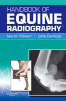
Handbook of Equine Radiography
W B Saunders Co Ltd (Verlag)
978-0-7020-2863-2 (ISBN)
- Titel ist leider vergriffen;
keine Neuauflage - Artikel merken
"The Handbook of Equine Radiography" is a practical and accessible 'how-to' guide to obtaining high-quality radiographs of the horse. It covers all aspects of taking radiographs of the commonly examined regions (lower limbs and skull) as well as less frequently examined areas (upper limbs, trunk). The main part of the book consists of diagrams to illustrate the positioning of the horse and the radiography equipment. For each view a high-quality digital example of a normal radiograph is illustrated. The accompanying text for each radiographic view succinctly presents the most relevant aspects. Practically orientated, and including chapters covering such key areas as radiation safety in equine radiography and patient preparation, the "Handbook of Equine Radiography" is an indispensable guide to practitioners in all countries engaged in equine work.
Part 1 Principles of radiography 1 Image formation 2 Radiographic equipment 3 Radiation safety and patient preparation 4 Radiological interpretation and diagnosis Part 2 Radiographic procedures 5 Radiography of the foot 6 Radiography of the pastern 7 Radiography of the fetlock 8 Radiography of the metacarpus and metatarsus 9 Radiography of the carpus 10 Radiography of the tarsus 11 Radiography of the elbow 12 Radiography of the shoulder 13 Radiography of the stifle 14 Radiography of the hip & pelvis 15 Radiography of the cervical vertebrae 16 Radiography of the thoracolumbar and sacral vertebrae 17 Radiography of the skull 18 Radiography of the thorax 19 Radiography of the abdomen 20 Additional radiographic procedures (contrast radiography) Appendix I Suggested exposure chart Appendix II Exposure chart for your practice Index
| Erscheint lt. Verlag | 28.11.2009 |
|---|---|
| Zusatzinfo | Approx. 230 illustrations (150 in full color) |
| Verlagsort | London |
| Sprache | englisch |
| Maße | 234 x 156 mm |
| Themenwelt | Veterinärmedizin ► Klinische Fächer ► Bildgebende Verfahren |
| Veterinärmedizin ► Pferd ► Bildgebende Verfahren | |
| ISBN-10 | 0-7020-2863-0 / 0702028630 |
| ISBN-13 | 978-0-7020-2863-2 / 9780702028632 |
| Zustand | Neuware |
| Haben Sie eine Frage zum Produkt? |
aus dem Bereich


