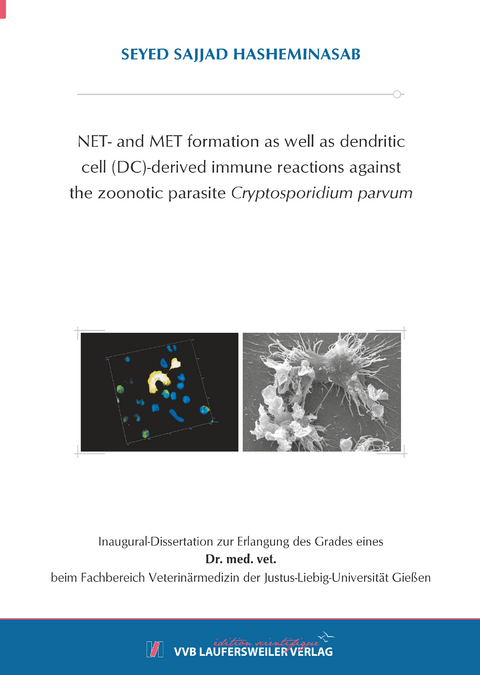
NET- and MET formation as well as dendritic cell (DC)-derived immune reactions against the zoonotic parasite Cryptosporidium parvum
Seiten
2024
VVB Laufersweiler Verlag
978-3-8359-7179-0 (ISBN)
VVB Laufersweiler Verlag
978-3-8359-7179-0 (ISBN)
Cryptosporidium parvum, ein protozoischer Parasit, ist weltweit verbreitet und kann sowohl Menschen als auch Nutztiere infizieren. Das Hauptreservoir für diese parasitäre Infektion sind neugeborene Kälber. Das Auftreten einer lebensbedrohlichen Kryptosporidiose bei HIV-Patienten, die mit C. parvum infiziert sind, macht deutlich, wie wichtig eine starke adaptive Immunreaktion für die wirksame Bekämpfung dieses Parasiten ist. Derzeit gibt es nur begrenzte Erkenntnisse über die Aktivierung der angeborenen und adaptiven Immunreaktion gegen C. parvum bei Menschen und Rindern. Neutrophil extracellular traps (NETs), die auch als suizidale NETosis bezeichnet werden, sind ein hochwirksamer und seit langem bestehender angeborener Abwehrmechanismus, der von polymorphkernigen Neutrophilen (PMN) zur Bekämpfung parasitärer Organismen wie Protozoen und Helminthen eingesetzt wird. Monozyten von Säugetieren sind eine Art von myeloischen Leukozyten, die aus dem Knochenmark stammen. Aufgrund ihrer verschiedenen Abwehrmechanismen spielen sie eine entscheidende Rolle bei der frühen angeborenen Immunantwort des Wirts. Dazu gehören das Vorhandensein von ATP-purinergen, CD14- und CD16-Rezeptoren sowie ihre Fähigkeit, an Oberflächen zu haften, zu wandern und Krankheitserreger zu phagozytieren. Monozyten besitzen auch entzündungshemmende und antiparasitäre Eigenschaften, was ihre wichtige Rolle im Immunsystem weiter unterstreicht. In jüngster Zeit wurde über die Bildung von extrazellulären Monozytenfallen (METs) als zusätzlicher Mechanismus zur Bekämpfung apikomplexer Parasiten berichtet. Allerdings gibt es in der Literatur derzeit keine Informationen über die Extrusion von METs in Bezug auf C. parvum-Oozysten oder Sporozoiten. In dieser Studie untersuchten wir den purinergen ATP-Rezeptor P2X1, die Glykolyse, das Notch-Signalisierung und die Laktat-Monocarboxylat-Transporter (MCT) in Rindermonozyten und PMN, die C. parvum unter zwei verschiedenen Sauerstoffbedingungen ausgesetzt waren: intestinale Physioxie (5 % O2) und Hyperoxie (21 % O2, was in Laborumgebungen üblich ist). Der Nachweis durch C. parvum ausgelöster suizidaler METs gelang durch die Anwendung mehrerer Verfahren. Erstens wurde mit Hilfe der Rasterelektronenmikroskopie eine vollständige Ruptur der exponierten Monozyten beobachtet. Darüber hinaus wurde die Co-Lokalisierung von extrazellulärer DNA mit Myeloperoxidase (MPO) und Histonen (H1-H4) Durch Analyse von Immunofluoreszenz- und Konfokalmikroskopieaufnahmen nachgewiesen. Nach rasterelektronenmikroskopischen (SEM) Analysen führten die von C. parvum induzierten suizidalen METs nicht nur zum Einschluss der Oozysten, sondern behindern das Austreten der Sporozoiten aus den Oozysten. Mit Hilfe der 3D-holotomographischen Analyse von lebenden Zellen konnten wir die frühe Aktivierung von Rindermonozyten durch die Parasiten dokumentieren. Diese Aktivierung war durch die Bildung von Membranausstülpungen bei Kontakt zu C. parvum. Die Analyse mit Hilfe der 3D-holotomographischen Mikroskopie von lebenden Zellen machte frühe morphologische Veränderungen in PMNs sichtbar, die durch Parasiten ausgelöst wurden. Zu diesen Veränderungen gehörte die Bildung von Membranausstülpungen in Richtung C. parvum während der NETose. Ein signifikanter Rückgang der durch C. parvum ausgelösten suizidal NETosis wurde beobachtet, wenn PMNs mit dem purinergen Rezeptor P2X1-Inhibitor NF449 behandelt wurden, unabhängig von den Sauerstoffbedingungen. Dieses Ergebnis deutet darauf hin, dass der P2X1-Rezeptor eine entscheidende Rolle bei der Bildung von NETs spielt, In ähnlicher Weise wurde bei der Hemmung der PMN-Glykolyse durch die Behandlung mit 2-Desoxy-Glukose ein leichter Rückgang der durch C. parvum ausgelösten suizidalen NETosis festgestellt, wenn auch nicht in signifikantem Ausmaß. Nach Messungen des energetischen Zustands der PMN führte die Exposition gegenüber C. parvum nicht zu einem Anstieg der extrazellulären Säuerungsrate (ECAR) oder der Sauerstoffverbrauchsrate (OCR) in den Zellen. Die Verwendung von Inhibitoren, die auf die Monocarboxylat-Transporter (MCTs) der Plasmamembran für Laktat abzielen, führte nicht zu einer signifikanten Verringerung der C. parvum-induzierten NET-Extrusion. Nach Behandlungen mit zwei spezifischen Notch-Inhibitoren (DAPT und Verbindung E), wurde in Bezug auf PMN keine signifikante Verringerung der Notch-Signalübertragung beobachtet. In dieser Studie beschreiben wir erstmals die wichtige Rolle des purinergen ATP-Rezeptors P2X1 bei der durch C. parvum ausgelösten suizidal NETosis unter Physioxie (5 % O2). Außerdem heben wir die antikryptosporidialen Eigenschaften dieses Rezeptors hervor. Wenn Monozyten verschiedenen Sauerstoffkonzentrationen ausgesetzt wurden, führte die Verabreichung von NF449, einem Inhibitor des purinergen ATP-Rezeptors P2X1, nicht zu einer signifikanten Verringerung der C. parvum-induzierten METosis. Dies deutet darauf hin, dass der Zelltodprozess nicht von P2X1 abhängt. Wenn Monozyten mit 2-Desoxy-Glukose (2-DG) behandelt wurden, um die Glykolyse zu hemmen, kam es jedoch zu einer Verringerung der durch C. parvum ausgelösten METosis, obwohl der Rückgang statistisch nicht signifikant war. Anhand von Messungen des energetischen Zustands der Monozyten wurde festgestellt, dass die Zellen, die C. parvum ausgesetzt waren, keinen Anstieg der extrazellulären Säuerungsrate (ECAR) oder der Sauerstoffverbrauchsrate (OCR) aufwiesen. Die Behandlung mit einem Inhibitor von Laktat-Monocarboxylat-Transportern (MCT), wie AR-C 141990, reduzierte die durch C. parvum induzierte Extrusion von METs unter physioxischen Bedingungen (5% O2) signifikant. Auch die Verabreichung von DAPT oder Compound E, zwei selektiven Notch-Inhibitoren, hatte keine nennenswerten hemmenden Auswirkungen auf die Produktion von Rinder-MET. In dieser Studie präsentieren wir den ersten Nachweis für die von C. parvum vermittelte METose als Abwehrmechanismus. Wir fanden heraus, dass dieser Prozess unabhängig von P2X1 ist, aber auf MCT beruht. Außerdem konnten wir beobachten, dass diese Effekte speziell unter intestinalen Physioxie-Bedingungen mit 5% CO2 auftreten. Die Ergebnisse der METs deuten darauf hin, dass es antikryptosporidiale Effekte gibt, die durch das Einfangen der Parasiten und die Hemmung des Prozesses der Sporozoiten-Exzystation erreicht werden.
Die Interaktion zwischen dendritischen Zellen (DC) und pathogenen Mikroorganismen ist ein entscheidender erster Schritt bei jeder adaptiven Immunantwort. Dendritische Zellen (DC) erkennen von Krankheitserregern stammende Moleküle und Alarmsignale und reagieren mit Aktivierung und Reifung. Wir haben primäre menschliche dendritische Zellen (DC) aus Monozyten von gesunden Blutspendern erzeugt. Diese DCs wurden dann in einer kontrollierten Laborumgebung C. parvum-Oozysten und Sporozoiten ausgesetzt. Im Allgemeinen erhöhte die Exposition gegenüber den Parasiten die Produktion der pro-inflammatorischen Zytokine/Chemokine IL-6 und IL-8 in den DC deutlich. Dieser Anstieg war vergleichbar mit dem Niveau, das bei der Stimulation mit Lipopolysaccharid (LPS), das als Positivkontrolle verwendet wurde, beobachtet wurde. Darüber hinaus kam es zu einer Hochregulierung von Reifungsmarkern und kostimulatorischen Molekülen, die für die Stimulation von T-Zellen notwendig sind, wie CD83, CD40 und CD86. Antigen-präsentierende Moleküle wie HLA-DR und CD1a sowie Adhäsionsmoleküle wie CD11b und CD58 wurden ebenfalls hochreguliert. Darüber hinaus zeigten menschliche DCs, die Parasiten ausgesetzt waren, eine verbesserte Zelladhäsion, eine erhöhte Mobilität und eine gesteigerte, wenn auch vorübergehende Fähigkeit, Oozysten und Sporozoiten von C. parvum zu zu phagozytieren. Diese verbesserte Phagozytose ist eine zusätzliche Voraussetzung für eine wirksame Antigenpräsentation. Im Gegensatz zu anderen mikrobiellen Stimuli führte die Exposition gegenüber C. parvum zu erhöhten oxidativen Verbrauchsraten (OCR) und nicht zu extrazellulären Säuerungsraten (ECAR) in DC. Dies deutet darauf hin, dass unterschiedliche Stoffwechselwege genutzt wurden, um Energie für die DC-Aktivierung zu erzeugen. Wenn menschliche DCs C. parvum ausgesetzt wurden, wiesen sie alle Merkmale einer erfolgreichen Reifung auf, die es ihnen ermöglichte, eine adaptive Immunantwort im Wirt wirksam zu initiieren. Cryptosporidium parvum, a zoonotic protozoan parasite, is found globally and has the ability to infect both humans and livestock. The main reservoir of this parasitic infection are neonatal calves. The occurrence of life-threatening cryptosporidiosis in HIV patients infected with C. parvum highlights the importance of a strong adaptive immune response in effectively combating this parasite. Currently, there is limited knowledge regarding the activation of innate and adaptive immune responses against C. parvum in humans and bovines. Neutrophil extracellular traps (NETs), also referred to as suicidal NETosis, are a highly effective and long-standing innate defense mechanism used by polymorphonuclear neutrophils (PMN) to combat parasitic organisms such as protozoa and helminths. Mammalian monocytes are a type of myeloid leukocytes that originate from the bone marrow. They play a crucial role in the early innate immune response of the host due to their various defense mechanisms. These include the presence of ATP purinergic-, CD14- and CD16-receptors, as well as their ability to adhere to surfaces, migrate, and phagocytose pathogens. Monocytes also possess inflammatory and anti-parasitic properties, further contributing to their important role in the immune system. Recently, there have been reports on the formation of monocyte extracellular traps (METs) as an additional mechanism to combat apicomplexan parasites. However, there is currently no information available in the literature regarding the extrusion of METs in relation to C. parvum-oocysts or sporozoites. In this study, we investigated the ATP purinergic receptor P2X1, glycolysis, Notch signaling, and lactate monocarboxylate transporters (MCT) in bovine monocytes and PMN exposed to C. parvum under two different oxygen conditions: intestinal physioxia (5% O2) and hyperoxia (21% O2, which is commonly used in laboratory settings). The confirmation of C. parvum-triggered suicidal NETs/METs were achieved through several methods. Firstly, the complete rupture of exposed monocytes and PMN were observed. Additionally, the co-localization of extracellular DNA with myeloperoxidase (MPO) and histones (H1-H4) was detected using immunofluorescence- and confocal microscopy analyses. According to scanning electron microscopy (SEM) analyses, suicidal NETs/METs induced by C. parvum not only led to oocyst entrapment but also hindered the egress of sporozoites from the oocysts. Using live cell 3D-holotomographic microscopy analysis, we were able to uncover the early activation of bovine PMN/monocytes induced by parasites. This activation was characterized by the formation of membrane protrusions towards C. parvum-oocysts/sporozoites.
The analysis using live cell 3D-holotomographic microscopy visualized early morphological changes in PMN induced by C. parvum. These changes included the formation of membrane protrusions towards C. parvum while undergoing NETosis. A significant decrease in C. parvum-induced suicidal NETosis was observed when PMN were treated with the purinergic receptor P2X1 inhibitor NF449, regardless of the oxygen conditions. This finding suggests that the P2X1 receptor plays a crucial role in the formation of NETs, highlighting its significance. In a similar way, when PMN glycolysis was inhibited through treatments with 2-deoxy glucose, there was a slight decrease in C. parvum-triggered suicidal NETosis, although not to a significant extent. According to measurements of PMN energetic state, the exposure to C. parvum did not result in an increase in extracellular acidification rates (ECAR) or oxygen consumption rates (OCR) in the cells. The use of inhibitors targeting plasma membrane monocarboxylate transporters (MCTs) of lactate resulted in a significant reduction of C. parvum-induced NET extrusion. No significant reduction in Notch signaling was observed following treatments with two specific Notch inhibitors, namely DAPT and compound E, in relation to PMN. In this study, we present the first description of the important role played by the ATP purinergic receptor P2X1 in C. parvum-induced suicidal NETosis under physioxia (5% O2). Additionally, we highlight the anti-cryptosporidial properties of this receptor. When monocytes were exposed to different levels of oxygen, the administration of NF449, an inhibitor of the ATP purinergic receptor P2X1, did not result in a significant reduction in C. parvum-induced METosis. This implies that the process of cell death does not rely on P2X1. Furthermore, when monocytes were treated with 2-deoxy glucose (2-DG) to inhibit glycolysis, there was a reduction in C. parvum-induced METosis, although the decrease was not statistically significant. Based on measurements of monocyte energetic state, it was found that cells exposed to C. parvum did not show any increase in extracellular acidification rates (ECAR) or oxygen consumption rates (OCR). The treatment with an inhibitor of lactate monocarboxylate transporters (MCT), such as AR-C 141990, significantly reduced the extrusion of METs induced by C. parvum under physioxic conditions (5% O2). In the same vein, the administration of either DAPT or compound E, which are two selective Notch inhibitors, did not show any notable inhibitory effects on the production of bovine MET. In this study, we present the first evidence of C. parvum-mediated METosis as a defense mechanism. We found that this process is independent of P2X1, but relies on MCT. Moreover, we observed that these effects occur specifically under intestinal physioxia conditions with 5% CO2. The findings from METs suggest that there are anti-cryptosporidial effects achieved by trapping the parasites and inhibiting the process of sporozoite excystation.
The interaction between dendritic cells (DC) and pathogenic microorganisms is a crucial first step in any adaptive immune response. DC detect molecules derived from pathogens and alarm signals, and respond by activating and maturing. We generated primary human DC from monocytes (MO-DC) obtained from healthy blood donors. These MO-DC were then exposed to C. parvum oocysts and sporozoites in a controlled laboratory environment. In general, exposure to parasites significantly increased the production of the pro-inflammatory cytokines/chemokines IL-6 and IL-8 in DC. This increase was comparable to the level observed with lipopolysaccharide (LPS) stimulation, which was used as a positive control. In addition, there was an upregulation of maturation markers and costimulatory molecules necessary for T cell stimulation, such as CD83, CD40, and CD86. Antigen-presenting molecules like HLA-DR and CD1a, as well as adhesion molecules like CD11b and CD58 were also upregulated. Furthermore, human DCs exposed to parasites exhibited improved cell adherence, increased mobility, and a heightened, albeit temporary, ability to engulf C. parvum oocysts and sporozoites. This enhanced phagocytosis serves as an additional requirement for effective antigen presentation. In contrast to other microbial stimuli, exposure to C. parvum resulted in increased oxidative consumption rates (OCR) rather than extracellular acidification rates (ECAR) in DC. This suggests that different metabolic pathways were utilized to generate energy for DC activation. When human DC were exposed to C. parvum, they exhibited all the characteristics of successful maturation, which allowed them to effectively initiate an adaptive immune response in the host.
| Erscheinungsdatum | 20.03.2024 |
|---|---|
| Reihe/Serie | Edition Scientifique |
| Verlagsort | Gießen |
| Sprache | englisch |
| Maße | 148 x 210 mm |
| Gewicht | 320 g |
| Themenwelt | Veterinärmedizin ► Allgemein |
| Schlagworte | Cryptosporidium parvum • HIV • Immunität • Immunreaktion • Kryptosporidiose • neugeborene Kälber • protozoischer Parasit |
| ISBN-10 | 3-8359-7179-4 / 3835971794 |
| ISBN-13 | 978-3-8359-7179-0 / 9783835971790 |
| Zustand | Neuware |
| Informationen gemäß Produktsicherheitsverordnung (GPSR) | |
| Haben Sie eine Frage zum Produkt? |

