Lipopolysaccharid-induzierte Toleranz und Sensibilisierung:
Effekte auf Primärkulturen des afferenten nozizeptiven Systems
Seiten
2021
VVB Laufersweiler Verlag
978-3-8359-6988-9 (ISBN)
VVB Laufersweiler Verlag
978-3-8359-6988-9 (ISBN)
- Keine Verlagsinformationen verfügbar
- Artikel merken
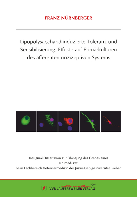
Eine der negativen Konsequenzen einer entzündlichen Stimulation des afferenten somatosensorischen Systems ist die Manifestation inflammatorischer Schmerzen. Um die hierfür verantwortlichen Mechanismen auf zellulärer Ebene untersuchen zu können, war das zentrale Ziel des ersten Teils der vorgelegten kumulativen Dissertationsschrift die Etablierung und Charakterisierung einer neuroglialen Zellkultur des dorsalen Anteils des Rückenmarks („superficial dorsal horn“, SDH), wo die afferenten Fasern der primären Nozizeptoren auf sekundäre Neurone verschaltet werden. Die SDH Kulturen setzen sich aus Neuronen (43 %), Oligodendrozyten (35 %), Astrozyten (13 %) und Mikroglia (9 %) zusammen. Von den SDH Neuronen reagierten 80 % auf Glutamat, 43 % auf Substanz P, 8 % auf Prostaglandin E2 (PGE2) und 100 % auf KCl (Vitalitätstest) mit einem Anstieg der intrazellulären Ca2+-Konzentration. Eine Kurzzeit-Stimulation mit 10 µg/ml Lipopolysaccharid (LPS, hohe Dosis) führte zu gesteigerter Expression von Zytokinen, inflammatorischen Transkriptionsfaktoren und induzierbaren Enzymen zur PGE2-Synthese. Auf Proteinebene wurden erhöhte Konzentrationen der Zytokine TNF-α und IL-6 in den Überständen LPS-stimulierter SDH Kulturen und erhöhte Immunreaktivität beider Zytokine in Mikrogliazellen nachgewiesen. Mikrogliazellen LPS-behandelter Kulturen zeigten außerdem verstärkte Immunreaktivität der inflammatorischen Transkriptionsfaktoren NFκB, NF-IL6 und pCREB in den Zellkernen. Nach Kurzzeit-Stimulation mit einer hohen LPS-Dosis war die Stärke der durch Substanz P induzierten Ca2+-Signale, nicht aber der Glutamat-induzierten Antworten, reduziert. Eine Langzeit-Stimulierung mit einer niedrigen LPS-Dosis (0.01 µg/ml für 24 Stunden) führte dagegen zu einer signifikanten Verstärkung der Glutamat-induzierten Ca2+-Signale, während die Antworten der Neurone auf Substanz P hierdurch unbeeinflusst blieben. Die Ergebnisse dieses Teils der Arbeit legen nahe, dass Mikrogliazellen im SDH des Rückenmarks entscheidend an der Ausbildung entzündlicher Prozesse beteiligt sind, die letztendlich zu veränderten neuronalen Reaktionsmustern führen.
Im zweiten Teil der vorgelegten Arbeit wurde experimentell untersucht, ob sich das Phänomen der „LPS-Toleranz“ in Strukturen des afferenten somatosensorischen Systems (Spinalganglien, „dorsal root ganglia“, DRG und SDH) manifestiert. Unter LPS-Toleranz versteht man die Abschwächung der inflammatorischen Zellantwort bei wiederholter Exposition. In beiden Primärkulturen konnten Effekte einer LPS-Toleranz durch Langzeit-Vorstimulation mit einer moderaten LPS-Dosis (1 µg/ml, 18 Stunden) gefolgt von einer Kurzzeit-Stimulation mit einer hohen Dosis (10 µg/ml, 2 Stunden) induziert werden. Auf mRNA Ebene war der LPS-tolerante Zustand besonders durch eine signifikante Abschwächung der Expression von TNF-α bzw. einem Anstieg des IL-10/TNF-α Expressionsverhältnisses charakterisiert. Auf Proteinebene war dies anhand reduzierter TNF-α Immunreaktivität in Makrophagen (DRG) und Mikrogliazellen (SDH) und verminderter Freisetzung von TNF-α in die Kulturüberstände (SDH) ebenfalls nachweisbar. Außerdem zeigten LPS-aktivierte inflammatorische Transkriptionsfaktoren im Zustand der LPS-Toleranz veränderte Aktivierungsmuster. Speziell die für die Expression inflammatorischer Mediatoren verantwortliche p65-Untereinheit von NFκB war in Makrophagen (DRG) und Mikrogliazellen (SDH) signifikant reduziert. Dies traf auch für den Transkriptionsfaktor NF-IL6 zu. Eine IL-6 vermittelte Aktivierung des Transkriptionsfaktors STAT3 war im LPS-toleranten Zustand in DRG-Neuronen und SDH-Astrozyten ebenfalls vermindert. Diese Ergebnisse zeigen, dass sich LPS-Toleranz in Strukturen des afferenten somatosensorischen Systems induzieren lässt und durch eine Abschwächung LPS-induzierter inflammatorischer Mediatoren und Transkriptionsfaktoren charakterisiert ist. Ein Einfluss der LPS-Toleranz auf neuronale Reaktivitätsmuster unter inflammatorischen Bedingungen bietet Möglichkeiten für weiterführende Untersuchungen.
Nach der experimentellen Untersuchung der LPS-Toleranz sollte im dritten Teil der vorgelegten Dissertationsschrift untersucht werden, ob sich auch das Phänomen einer „LPS-Sensibilisierung“ in Primärkulturen des afferenten somatosensorischen Systems (hier: Spinalganglien, DRG) nachweisen lässt. Hierbei handelt es sich um einen bislang postulierten, aber kaum nachgewiesenen Zustand, in dem eine Vorbehandlung mit sehr niedrigen LPS-Dosierungen ohne Eigeneffekt zu einer verstärkten Reaktion auf Stimulierung mit einer hohen LPS-Dosis führt. Von zahlreichen getesteten LPS-Dosierungen konnte eine starke Tendenz für einen Sensibilisierungseffekt für die Dosis von 0.001 µg/ml LPS für 18 Stunden anhand der TNF-α Freisetzung identifiziert werden. Diese und weitere unterschwellige LPS-Dosierungen waren jedoch nicht in der Lage, Capsaicin-induzierte Ca2+-Signale in primären nozizeptiven Neuronen zu verstärken. Eine inflammatorische Verstärkung Capsaicin-induzierter neuronaler Ca2+-Signale war nur durch Vorbehandlung mit einer LPS-Dosis (1 µg/ml für 18 Stunden) möglich, die per se auch zur deutlichen Freisetzung von Zytokinen (TNF-α) in die Kulturüberstände führt.
Die Ergebnisse dieser Arbeit zeigen, dass DRG und SDH Primärzellkulturen gut als Experimentalmodelle geeignet sind, um den Einfluss inflammatorischer Stimuli auf das afferente somatosensorische System zu untersuchen. Speziell für LPS können auch Zustände der LPS-Toleranz und der LPS-Sensibilisierung mit Hilfe der Kulturen untersucht und charakterisiert werden. Perspektivisch könnten beide Kulturen gemäß dem 3R Prinzip für Screenings von Substanzen mit potentiellen anti-inflammatorischen bzw. anti-nozizeptiven Eigenschaften herangezogen werden. One maladaptive consequence of inflammatory stimulation of the afferent somatosensory system is the manifestation of inflammatory pain. The central goal of the first part of this thesis was to establish and characterize a neuroglial primary culture of the rat superficial dorsal horn (SDH) of the spinal cord, mainly including the substantia gelatinosa, to test responses of this structure to neurochemical, somatosensory, or inflammatory stimulation. Primary cultures of the rat SDH consist of neurons (43 %), oligodendrocytes (35 %), astrocytes (13 %) and microglial cells (9 %). Neurons of the SDH responded to cooling (7 %), heating (18 %), glutamate (80 %), substance P (43 %), prostaglandin E2 (8 %) and KCl (100 %) with transient increases in the intracellular calcium [Ca2+]i. Short-term stimulation of SDH primary cultures with lipopolysaccharide (LPS, 10 µg/ml, 2 h) caused increased expression of pro-inflammatory cytokines, inflammatory transcription factors and inducible enzymes responsible for inflammatory prostaglandin E2-synthesis. At the protein level, increased concentrations of tumor necrosis factor-α (TNF-α) and interleukin-6 (IL-6) were measured in the supernatants of LPS-stimulated SDH cultures and enhanced TNF-α and IL-6 immunoreactivity was observed specifically in microglial cells. LPS-exposed microglial cells further showed increased nuclear immunoreactivity for the inflammatory transcription factors NFκB, NF-IL6 and pCREB, indicative for their activation. The short-term exposure to LPS further caused a reduction in the strength of substance P- as opposed to glutamate-evoked Ca2+-signals in SDH neurons. However, long-term stimulation with a low dose of LPS (0.01 μg/ml, 24 h) resulted in a significant increase of glutamate-induced Ca2+-transients in SDH neurons, while substance P-evoked Ca2+-signals were not influenced. Our data suggest a critical role for microglial cells in the initiation of inflammatory processes within the SDH of the spinal cord, which are accompanied by a modulation of neuronal responses.
LPS can induce a state of refractoriness to its own effects termed LPS-tolerance. In the second part of this thesis we therefore employed primary neuroglial cultures from rat dorsal root ganglia (DRG) and the SDH of the spinal cord to establish and characterize a model of LPS-tolerance within these structures. Tolerance was induced by pre-treatment of both cultures with 1 µg/ml LPS for 18 h, followed by a short-term stimulation with a higher LPS-dose (10 µg/ml for 2 h). Cultures treated with solvent were used as controls. Cells from DRG or SDH were investigated by means of RT-PCR (expression of inflammatory genes) and immunocytochemistry (translocation of inflammatory transcription factors into nuclei of cells from both cultures). Supernatants from both cultures were assayed for tumor necrosis factor-α (TNF-α) and interleukin-6 (IL-6) by highly sensitive bioassays. At the mRNA-level, pre-treatment with 1 µg/ml LPS caused reduced expression of TNF-α and enhanced IL-10/TNF-α expression ratios in both cultures upon subsequent stimulation with 10 µg/ml LPS, i.e. LPS-tolerance. SDH cultures further showed reduced release of TNF-α into the supernatants and attenuated TNF-α-immunoreactivity in microglial cells. In the state of LPS-tolerance macrophages from DRG and microglial cells from SDH showed reduced LPS-induced nuclear translocation of the inflammatory transcription factors NFB and NF-IL6. Nuclear immunoreactivity of the IL-6-activated transcription factor STAT3 was further reduced in neurons from DRG and astrocytes from SDH in LPS-tolerant cultures. A state of LPS-tolerance can thus be induced in primary cultures from the afferent somatosensory system, which is characterized by a down-regulation of pro-inflammatory mediators. Furthermore, this model can be applied to study the effects of LPS-tolerance at the cellular level, for example possible modifications of neuronal reactivity patterns upon inflammatory stimulation.
Aim of the third part of the thesis was to investigate whether an exposure of mixed neuroglial primary cultures from dorsal root ganglia (DRG) with very low doses of LPS causes a state of “LPS-sensitization”. We tested whether priming of DRG cultures with ultra-low LPS-doses modified the TNF-α-release by the cells to a subsequent challenge with a high LPS-dose. We further investigated the capsaicin-evoked Ca2+-signals in neurons from DRG, which were pre-treated with a wide range of LPS-doses. Release of TNF-α into the supernatants was hardly modified by pre-exposure to low LPS-doses. Capsaicin-evoked Ca2+-signals were significantly enhanced by pre-treatment with LPS in a dose-dependent manner. Only a dose of LPS, which caused pronounced release of TNF-α into the supernatant, was capable to enhance the Ca2+-responses of putative nociceptors and to increase the sensitivity of these neurons to lower doses of capsaicin. We conclude that ultra-low doses of LPS, which per se do not evoke a detectable inflammatory response, are not sufficient to sensitize neurons (Ca2+-responses) and glial elements (TNF-α-responses) of the primary afferent somatosensory system.
In summary, mixed neuroglial primary cultures from DRG and SDH are useful tools to study the impact of experimentally induced inflammation on the afferent somatosensory system. With regard to LPS as inflammatory stimulus, specific conditions including LPS-tolerance or LPS-induced sensitization can be investigated and characterized. As a perspective both cultures may be used for screening of drugs with putative anti-inflammatory and/or anti-nociceptive properties
| Erscheinungsdatum | 10.01.2022 |
|---|---|
| Reihe/Serie | Edition Scientifique |
| Verlagsort | Gießen |
| Sprache | deutsch |
| Maße | 148 x 210 mm |
| Gewicht | 280 g |
| Themenwelt | Veterinärmedizin ► Allgemein |
| Schlagworte | Nerven • Neurologie • Schmerz |
| ISBN-10 | 3-8359-6988-9 / 3835969889 |
| ISBN-13 | 978-3-8359-6988-9 / 9783835969889 |
| Zustand | Neuware |
| Haben Sie eine Frage zum Produkt? |

