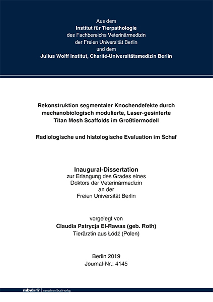
Rekonstruktion segmentaler Knochendefekte durch mechanobiologisch modulierte, Laser-gesinterte Titan Mesh Scaffolds im Großtiermodell - Radiologische und histologische Evaluation im Schaf
Seiten
2019
|
1. Aufl.
Mensch & Buch (Verlag)
978-3-86387-988-4 (ISBN)
Mensch & Buch (Verlag)
978-3-86387-988-4 (ISBN)
- Keine Verlagsinformationen verfügbar
- Artikel merken
Die Behandlung segmentaler Knochendefekte kritischer Größe, die durch Traumata, Tumorresektion oder Infektionen hervorgerufen werden, sowie die mechanobiologische Wiederherstellung der Gliedmaße, stellen nach wie vor eine Herausforderung in der Unfallchirurgie dar. Aufgrund dessen werden alternative Behandlungsmethoden von kritischen Knochendefekten zunehmend erforscht. Mit Hilfe der Laser-Sinterungstechnik hergestellte dreidimensionale Titan Mesh Scaffolds stellen eine vorteilhafte Alternativmethode zu den bekannten ,,Gold Standards“ dar und ließen beim klinischen Einsatz am humanen Patienten vielversprechende Rückschlüsse zu. In der vorliegenden Studie wurden mittels der Laser-Sinterungstechnik zwei Titan Mesh Scaffolds hergestellt, die strukturell gleich waren, sich jedoch in ihren Steifigkeiten voneinander unterschieden. Die Titan Mesh Scaffolds besaßen eine Honigwabenform und bildeten ein makro-poröses Netzwerk mit einer zentralen Pore. Durch die Veränderung des Strebendurchmessers der Titan Strut Elemente, entstanden zwei Titan Mesh Scaffolds mit unterschiedlichen Steifigkeiten. In der vorliegenden Studie erfolgte die Durchführung der 40 mm großen Osteotomie an der Tibia von zwölf Merino-Mix-Schafen. Es wurden zwei Gruppen mit jeweils sechs Tieren gebildet. In der einen Gruppe wurde der weiche Titan Mesh Scaffold mit einem Strebendiameter von 1.2 mm mit 0,84 GPA eingesetzt und bei der anderen Gruppe der 3,5-fach steife Titan Mesh Scaffold mit einem Strebendiameter von 1.6 mm mit 2,88 GPa eingesetzt. Die mit autologer Spongiosa befüllten Titan Mesh Scaffolds wurden in Kombination mit einem experimentellen winkelstabilen Plattensystem (AO-Platte), welches ausschließlich eine axiale Belastung der Gliedmaße zuließ, in den kritischen Osteotomiedefekt von 40 mm Größe in die Schafstibia eingesetzt. Während der Versuchszeit von 24 Wochen wurden monatliche Röntgenkontrollen durchgeführt. In der 24. Woche wurden ex vivo zusätzlich zu den konventionellen Röntgenaufnahmen, Aufnahmen im Faxitron angefertigt. Histologische und histomorphometrische Untersuchungsergebnisse wurden erfasst und evaluiert.
Der Einsatz des weichen und des 3,5-fach steiferen Titan Mesh Scaffolds in Kombination mit der experimentellen winkelstabilen AO-Platte erwies sich als eine adäquate Stabilisierungsmethode für einen kritischen Defekt von 40 mm Größe im Schafmodel. Die Hypothese, dass eine mechanisch-biologische Optimierung des Titan Mesh Scaffolds zu einer Förderung der endogenen Knochendefektregeneration führt, konnte histomorphologisch vermutet werden, da der Einsatz des weicheren Titan Mesh Scaffolds deskriptiv zu einer vermehrten Kallusbildung im Vergleich zu dem steiferen Titan Mesh Scaffold, zu beobachten war. Das Ergebnis konnte histomorphometrisch nicht bestätigt werden, da im Vergleich der beiden Gruppen kein statitisch signifikanter Unterschied vorlag.
Die AO-Platte wurde speziell für das Schaf entwickelt, stabilisierte den Fakturspalt ohne den Einsatz weiterer Stabilisierungsverfahren und gewährleistete zusätzlich eine artgerechte Haltung der Schafe. Die Titan Mesh Scaffolds füllten den Defekt gut aus und erwiesen sich als stabil, sie verhinderten einen Muskel- und Weichteilprolaps in den Defekt und dienten darüberhinaus als Leitstruktur für das wachsende Gewebe. Da nach 24 Wochen keine komplette Überbrückung des Defektspaltes zu beobachten war, lag eine verzögerte Heilung vor. Die mechanische Belastung im Frakturspalt wurde durch die Titan Mesh Scaffolds in Kombination mit der AO-Platte minimiert, sodass die Kallusformatinon gering war. Dennoch zeigte die Gewebezusammensetzung eine noch aktive Knochenheilung. Die in dieser Studie gewonnenen Erkenntnisse können genutzt werden, um die Kombination des Titan Mesh Scaffolds mit einer anderen Platte, die mehr mechanische Bewegung im Frakturspalt ermöglicht und demnach zu einer vermehrten Kallusbildung führen könnte, zu untersuchen. Reconstruction of large segmental bone defects with mechano-biologically optimized laser-sintered titanium mesh scaffolds in a translational large animal study
The treatment of segmental bone defects of critical size caused by trauma, tumor resection or infection, as well as the mechano-biological restoration of the limb, still pose a challenge in traumatology. As a result, alternative treatments for critical bone defects are increasingly being explored. Three-dimensional (3D) titanium mesh scaffolds produced by using finite element techniques represent an advantageous alternative method to the known "gold standards" and have given promising conclusions in clinical use on human patients. In our study, two titanium mesh scaffolds, with the same structure but different stiffness, were produced and inserted in a critical size bone defect model in vivo in sheep. In the present study, the 40 mm osteotomy was performed on the tibia of twelve merino-mix sheep. The titanium mesh scaffolds had honeycomb-like and a macro-porous network with a central pore. The stiffness was altered through small changes in the strut diameter. Two groups of six animals were formed. One group received the soft scaffold (0.84 GPa stiffness) and the other group received the 3.5-fold stiffer scaffold (2.88 GPa). The scaffolds of both groups were filled with the same amount of autologous cancellous bone graft (ABG; 7.5 ml) and were applied within the 40 mm defect. The defect was stabilized with a rigid custom made half shell steel plate (AO Research Institute Davos), which mechanically shielded the defect and allowed only an axial load on the limb. During the trial period of 24 weeks, monthly X-ray checks were performed. In the 24th week ex vivo, in addition to the conventional X-rays, Faxitron shots were taken. Histological and histomorphometric test results were recorded and evaluated. The use of the soft and the 3.5 times stiffer titanium mesh scaffold in combination with a rigid, custom-made shielding plate proved to be an adequate treatment method for a critical defect of 40 mm in the sheep model. The hypothesis that mechanicalbiological optimization of the titanium mesh scaffold promotes endogenous bone defect regeneration could be assumed histomorphologically, as the use of the softer titanium mesh scaffold resulted in an increased callus formation compared to the more rigid titanium mesh scaffold. The result could not be confirmed histomorphometrically, the comparison of both groups showed no difference in the statistical evaluation. The rigid, custom-made shielding plate was specially designed for sheep. It stabilized the fracture gap without the need for other stabilization methods and allowing standard animal welfare. The titanium mesh scaffold filled out the fracture gap and proved to be a stable method. It prevented a prolapse of muscle and soft tissue into the gap and turned out to be a lead structure for the ingrowing bone. Since a complete bridging could not be seen after 24 weeks delayed healing could be identified. The titanium mesh scaffold in combination with the rigid, custom-made shielding plate reduced the mechanical load in the fracture gap, so that the callus formation was limited, but still active. The results of this study can be used as a basic method for further research of a titanium mesh scaffold in combination with another plate system, which allows more mechanical load into the gap and leads to an increased callus formation.
| Erscheinungsdatum | 20.09.2019 |
|---|---|
| Verlagsort | Berlin |
| Sprache | deutsch |
| Maße | 148 x 210 mm |
| Themenwelt | Veterinärmedizin ► Vorklinik ► Histologie / Embryologie |
| Veterinärmedizin ► Klinische Fächer ► Bildgebende Verfahren | |
| Veterinärmedizin ► Großtier ► Schaf / Ziege | |
| Schlagworte | Animal Models • bone fractures • chirurgische Eingriffe • Clinical Trials • Fracture Fixation • Frakturfixation • Großtiermodell • Histologie • Histology • Implantation • klinische Versuche • Knochenbrüche • Knochendefekte • Schaf • Sheep • surgical operations • Tiermodell • Titan Mesh Scaffolds |
| ISBN-10 | 3-86387-988-0 / 3863879880 |
| ISBN-13 | 978-3-86387-988-4 / 9783863879884 |
| Zustand | Neuware |
| Informationen gemäß Produktsicherheitsverordnung (GPSR) | |
| Haben Sie eine Frage zum Produkt? |
Mehr entdecken
aus dem Bereich
aus dem Bereich
Differentialdiagnosen beim Kleintier
Buch | Softcover (2023)
LABOKLIN GmbH & Co. KG (Verlag)
55,95 €
Grundlagen, Techniken, Präparate
Buch | Hardcover (2019)
Schlütersche (Verlag)
59,95 €


