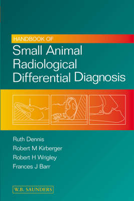
Handbook of Small Animal Radiological Differential Diagnosis
Seiten
2001
W B Saunders Co Ltd (Verlag)
978-0-7020-2485-6 (ISBN)
W B Saunders Co Ltd (Verlag)
978-0-7020-2485-6 (ISBN)
- Titel erscheint in neuer Auflage
- Artikel merken
Zu diesem Artikel existiert eine Nachauflage
Intended as an aide memoir of differential diagnoses, and other useful information in small animal radiology and ultrasound, in order to assist the radiologist to compile as a list of differential diagnoses. This book is useful for users of small animal diagnostic imaging, from radiologists through general practitioners to veterinary students.
This book is the veterinary equivalent of the popular medical book "Aids to Radiological Differential Diagnosis" by Chapman and Nakielny, and will prove invaluable to all users of small animal diagnostic imaging, from radiologists through general practitioners to veterinary students. It is intended as an aide memoire of differential diagnoses and much other useful information in small animal radiology and ultrasound, in order to assist the radiologist to compile as complete a list of differential diagnoses as possible. Some details of radiographic technique (including contrast studies) are included and guidance on ultrasonographic technique and a description of the normal ultrasonographic appearance of organs is given. A large number of clear, schematic line drawings of many of the conditions are included, to complement the text. The book is divided into 12 chapters each representing different body systems, and for various radiographic and ultrasonographic abnormalities possible diagnoses are listed in approximate order of likelihood, including those due to normal anatomical variation and technical or iatrogenic causes.
Conditions which principally or exclusively occur in cats are indicated, although many of the other diseases listed may occur in cats as well as in dogs. Infectious and parasitic diseases which are not ubiquitous but which are confined to certain parts of the world are indicated as such, and a comprehensive table in the Appendix lists their geographic distribution. The Appendix also includes useful at-a-glance summaries of Radiographic Faults and Ultrasound Terminology and Artefacts. Lists of references for further reading appear at the end of each chapter and these will prove helpful to the reader seeking further information about a particular condition.
Preface. Foreword. 1. Skeletal system: general. 2. Joints. 3. Appendicular skeleton. 4. Head and neck. 5. Spine. 6. Lower respiratory tract. 7. Cardiovascular system. 8. Other thoracic structures - pleural cavity, mediastinum thoracic oesophagus, thoracic wall. 9. Gastrointestinal tract. 10. Urogenital tract. 11. Other abdominal structures - abdominal wall, peritoneal and retroperitoneal cavities, parenchymal organs. 12. Soft tissues. Appendix. Index.
| Erscheint lt. Verlag | 21.5.2001 |
|---|---|
| Zusatzinfo | 280 ills. |
| Verlagsort | London |
| Sprache | englisch |
| Maße | 156 x 234 mm |
| Gewicht | 485 g |
| Themenwelt | Veterinärmedizin ► Kleintier |
| ISBN-10 | 0-7020-2485-6 / 0702024856 |
| ISBN-13 | 978-0-7020-2485-6 / 9780702024856 |
| Zustand | Neuware |
| Haben Sie eine Frage zum Produkt? |
Mehr entdecken
aus dem Bereich
aus dem Bereich
Clinical Medicine and Management
Buch | Hardcover (2024)
Elsevier - Health Sciences Division (Verlag)
199,95 €
Blackwell's Five-Minute Veterinary Consult
Buch | Hardcover (2021)
Wiley-Blackwell (Verlag)
115,00 €


