Handbook of Nuclear Medicine and Molecular Imaging for Physicists
CRC Press (Verlag)
978-1-138-59329-9 (ISBN)
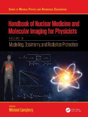
Mathematical modelling is an important part of nuclear medicine. Therefore, several chapters of this book have been dedicated towards describing this topic. In these chapters, an emphasis has been put on describing the mathematical modelling of the radiation transport of photons and electrons, as well as on the transportation of radiopharmaceuticals between different organs and compartments. It also includes computer models of patient dosimetry. Two chapters of this book are devoted towards introducing the concept of biostatistics and radiobiology. These chapters are followed by chapters detailing dosimetry procedures commonly used in the context of diagnostic imaging, as well as patient-specific dosimetry for radiotherapy treatments.
For safety reasons, many of the methods used in nuclear medicine and molecular imaging are tightly regulated. Therefore, this volume also highlights the basic principles for radiation protection. It discusses the process of how guidelines and regulations aimed at minimizing radiation exposure are determined and implemented by international organisations. Finally, this book describes how different dosimetry methods may be utilized depending on the intended target, including whole-body or organ-specific imaging, as well as small-scale to cellular dosimetry.
This text will be an invaluable resource for libraries, institutions, and clinical and academic medical physicists searching for a complete account of what defines nuclear medicine.
The most comprehensive reference available providing a state-of-the-art overview of the field of nuclear medicine
Edited by a leader in the field, with contributions from a team of experienced medical physicists, chemists, engineers, scientists, and clinical medical personnel
Includes the latest practical research in the field, in addition to explaining fundamental theory and the field's history
Michael Ljungberg is a Professor at Medical Radiation Physics, Lund, Lund University, Sweden. He started his research in the Monte Carlo field in 1983 through a project involving a simulation of whole-body counters but later changed the focus to more general applications in nuclear medicine imaging and SPECT. Parallel to his development of the Monte Carlo code, SIMIND, he began working in 1985 with quantitative SPECT and problems related to attenuation and scatter. After earning his PhD in 1990, he received a research assistant position that allowed him to continue developing SIMIND for quantitative SPECT applications and to establish successful collaborations with international research groups. At this time, the SIMIND program became used world-wide. Dr. Ljungberg became an associate professor in 1994 and, in 2005, after working clinically as a nuclear medicine medical physicist, received a full professorship in the Science Faculty at Lund University. He became the Head of the Department of Medical Radiation Physics at Lund in 2013 and a full professor in the Medical Faculty in 2015. Aside from the development of SIMIND – including new camera systems such as CZT detectors – his research includes an extensive project in oncological nuclear medicine. In this project, he and colleagues developed dosimetry methods based on quantitative SPECT, Monte Carlo absorbed-dose calculations, and methods for accurate 3D dose planning for internal radionuclide therapy. Lately, his work has focused on implementing Monte Carlo–based image reconstruction in SIMIND. He is also involved in the undergraduate education of medical physicists and bio-medical engineers and supervises MSc and PhD students. In 2012, Professor Ljungberg became a member of the European Association of Nuclear Medicines task group on Dosimetry and served that association for six years. He has published over a hundred original papers, 18 conference proceedings, 18 books and book chapters, and 14 peer-reviewed papers.
Contents
Preface...............................................................................................................................vii
Editor..................................................................................................................................ix
Contributors.......................................................................................................................xi
Chapter 1 Introduction to Biostatistics..........................................................................1
Johan Gustafsson and Markus Nilsson
Chapter 2 Radiobiology....................................................................................................17
Lidia Strigari and Marta Cremonesi
Chapter 3 Diagnostic Dosimetry.............................................................................................33
Lennart Johansson† and Martin Andersson
Chapter 4 Time- activity Curves: Data, Models, Curve Fitting, and Model Selection..........................69
Gerhard Glatting
Chapter 5 Tracer Kinetic Modelling and Its Use in PET Quantification..............................................83
Mark Lubberink and Michel Koole
Chapter 6 Principles of Radiological Protection in Healthcare..........................................................101
Soren Mattsson
Chapter 7 Controversies in Nuclear Medicine Dosimetry..................................................................115
Michael G. Stabin
Chapter 8 Monte Carlo Simulation of Photon and Electron Transport in Matter..............................123
Jose M. Fernandez-Varea
Chapter 9 Patient Models for Dosimetry Applications..........................................................141
Michael G. Stabin
Chapter 10 Patient- specific Dosimetry Calculations.............................................................155
Manuel Bardies, Naomi Clayton, Gunjan Kayal, and Alex Vergara Gil
Chapter 11 Whole- body Dosimetry..................................................169
Jonathan Gear
Chapter 12 Personalized Dosimetry in Radioembolization........................................................................................183
Remco Bastiaannet and Hugo W.A.M. de Jong
Chapter 13 Thyroid Imaging and Dosimetry..........................................................................207
Michael Lassmann and Heribert Hanscheid
Chapter 14 Bone Marrow Dosimetry........................................................................................223
Cecilia Hindorf
Chapter 15 Cellular and Multicellular Dosimetry...................................................................235
Roger W. Howell
Chapter 16 Alpha- particle Dosimetry................................................................267
Stig Palm
Chapter 17 Staff Radiation Protection........................................................................275
Lena Jonsson
Chapter 18 IAEA Support to Nuclear Medicine...........................................................293
Gian Luca Poli
| Erscheinungsdatum | 10.02.2022 |
|---|---|
| Reihe/Serie | Series in Medical Physics and Biomedical Engineering |
| Zusatzinfo | 29 Tables, black and white; 84 Line drawings, black and white; 57 Halftones, black and white; 141 Illustrations, black and white |
| Verlagsort | London |
| Sprache | englisch |
| Maße | 210 x 280 mm |
| Gewicht | 1320 g |
| Themenwelt | Informatik ► Grafik / Design ► Digitale Bildverarbeitung |
| Medizinische Fachgebiete ► Radiologie / Bildgebende Verfahren ► Nuklearmedizin | |
| Medizinische Fachgebiete ► Radiologie / Bildgebende Verfahren ► Radiologie | |
| Naturwissenschaften ► Physik / Astronomie ► Angewandte Physik | |
| Technik ► Umwelttechnik / Biotechnologie | |
| ISBN-10 | 1-138-59329-X / 113859329X |
| ISBN-13 | 978-1-138-59329-9 / 9781138593299 |
| Zustand | Neuware |
| Haben Sie eine Frage zum Produkt? |
aus dem Bereich


