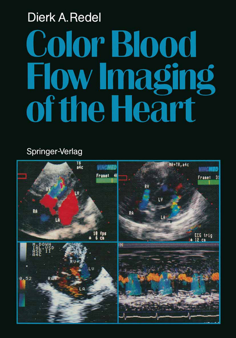
Color Blood Flow Imaging of the Heart
Springer Berlin (Verlag)
978-3-642-71174-9 (ISBN)
Preface.- 1 Introduction.- 2 Principles of Color Blood Flow Imaging.- 2.1 Acquisition of Blood Flow Data.- 2.2 Color Encoding.- 2.3 Color Aliasing.- 2.4 Sensitivity of CBFI.- 3 Artefacts in Color Blood Flow Imaging.- 3.1 Reverberations.- 3.2 Mirroring.- 3.3 Wall Motion Artefacts.- 3.4 Inappropriate Transducer Position.- 3.5 Distortion of Flow Velocity Display.- 3.6 Azimuth Artefacts.- 4 Color Display of Blood Flow Velocities.- 4.1 Physiological Flow with Flat Profile.- 4.2 Physiological Flow with Non-Flat Profile.- 4.3 Convective Acceleration.- 4.4 Streamline Separation.- 4.5 Angle Dependency of CBFI.- 4.6 Disturbed Flow.- 4.7 Differentiation of Flow Velocities from Wall Motion.- 5 Physiological Flow Velocity Patterns in the Cardiovascular System.- 5.1 Left Atrium and Pulmonary Veins.- 5.2 Mitral Valve.- 5.3 Left Ventricle.- 5.4 Aorta (Ascending, Arch, Descending).- 5.5 Right Atrium and Caval Veins.- 5.6 Tricuspid Valve.- 5.7 Right Ventricle.- 5.8 Pulmonary Artery and Main Branches.- 6 Blood Flow Velocity Patterns in Heart Disease.- 6.1 Shunts at or Above Atrial Level (Pretricuspid Shunts).- 6.2 Shunts at Ventricular Level (Post-tricuspid Shunts-1 -).- 6.3 Shunts at Arterial Level (Post-tricuspid Shunts - 2 -).- 6.4 Abnormalities of the Left Ventricular Inflow.- 6.5 Abnormalities of the Left Ventricular Outflow and the Aorta.- 6.6 Abnormalities of the Right Ventricular Inflow.- 6.7 Abnormalities of the Right Ventricular Outflow.- References.
| Erscheint lt. Verlag | 17.11.2011 |
|---|---|
| Zusatzinfo | VII, 130 p. |
| Verlagsort | Berlin |
| Sprache | englisch |
| Maße | 170 x 244 mm |
| Gewicht | 261 g |
| Themenwelt | Medizinische Fachgebiete ► Innere Medizin ► Kardiologie / Angiologie |
| Medizinische Fachgebiete ► Radiologie / Bildgebende Verfahren ► Sonographie / Echokardiographie | |
| Technik | |
| Schlagworte | Cardiology • Cardiovascular • Cardiovascular System • heart • Heart disease • Imaging • ultrasonography |
| ISBN-10 | 3-642-71174-X / 364271174X |
| ISBN-13 | 978-3-642-71174-9 / 9783642711749 |
| Zustand | Neuware |
| Haben Sie eine Frage zum Produkt? |
aus dem Bereich


