Surface Plasmon Enhanced, Coupled and Controlled Fluorescence
John Wiley & Sons Inc (Verlag)
978-1-118-02793-6 (ISBN)
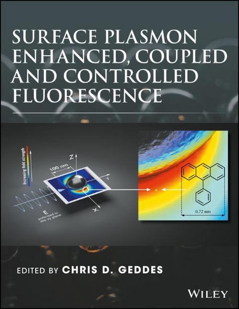
Explains the principles and current thinking behind plasmon enhanced Fluorescence
Describes the current developments in Surface Plasmon Enhanced, Coupled and Controlled Fluorescence
Details methods used to understand solar energy conversion, detect and quantify DNA more quickly and accurately, and enhance the timeliness and accuracy of digital immunoassays
Contains contributions by the world’s leading scientists in the area of fluorescence and plasmonics
Describes detailed experimental procedures for developing both surfaces and nanoparticles for applications in metal-enhanced fluorescence
Chris D. Geddes, PhD, FRSC, is a professor at the University of Maryland, Baltimore County, USA, where he is the director of the Institute of Fluorescence, and the editor-in-chief of both the Journal of Fluorescence and the Plasmonics journal. With more than 250 papers, 35 books, and >100 patents to his credit, he has extensive expertise in fluorescence spectroscopy, particularly in fluorescence sensing and metal–fluorophore interactions.
List of Contributors xi
Preface xv
1 Plasmonic–Fluorescent and Magnetic–Fluorescent Composite Nanoparticle as Multifunctional Cellular Probe 1
Arindam Saha, SK Basiruddin, and Nikhil Ranjan Jana
1.1 Introduction 1
1.2 Synthesis Design of Composite Nanoparticle 2
1.2.1 Method 1: Polyacrylate Coating–Based Composite of Nanoparticle and Organic Dye 3
1.2.2 Method 2: Polyacrylate Coating–Based Composite of Two Different Nanoparticles 3
1.2.3 Method 3: Ligand Exchange Approach–Based Composite of Two Different Nanoparticles 4
1.3 Property of Composite Nanoparticles 5
1.3.1 Optical Property 5
1.3.2 Fluorophore Lifetime Study 7
1.4 Functionalization and Labeling Application of Composite Nanoparticle 8
1.5 Conclusion 8
2 Compatibility of Metal–Induced Fluorescence Enhancement with Applications in Analytical Chemistry and Biosensing 13
Fang Xie, Wei Deng, and Ewa M. Goldys
2.1 Introduction 13
2.2 Homogeneous Protein Sensing MIFE Substrates 14
2.2.1 Core–Shell Approach 14
2.2.2 Homogeneous Large Au Nanoparticle Substrates 16
2.2.3 Commercial Klarite™ Substrate 18
2.3 Ag Fractal Structures 19
2.3.1 Reasons for High Enhancement Factors in Nanowire Structures 19
2.3.2 Ag Dendritic Structure—Homogeneous Silver Fractal 22
2.4 MIFE with Membranes for Protein Dot Blots 25
2.5 MIFE with Flow Cytometry Beads and Single Particle Imaging 30
3 Plasmonic Enhancement of Molecule–Doped Core–Shell and Nanoshell on Molecular Fluorescence 37
Jiunn–Woei Liaw, Chuan–Li Liu, Chong–Yu Jiang, and Mao–Kuen Kuo
3.1 Introduction 37
3.2 Theory 38
3.2.1 Plane Wave Interacting with an Multilayered Sphere 39
3.2.2 Excited Dipole Interacting with a Multilayered Sphere 40
3.2.3 EF on Fluorescence 40
3.3 Numerical Results and Discussion 41
3.3.1 Core–Shell 41
3.3.2 Nanoshelled Nanocavity 50
3.3.3 NS@SiO2 53
3.4 Conclusion 66
4 Controlling Metal–Enhanced Fluorescence Using Bimetallic Nanoparticles 73
Debosruti Dutta, Sanchari Chowdhury, Chi Ta Yang, Venkat R. Bhethanabotla, and Babu Joseph
4.1 Introduction 73
4.2 Experimental Methods 74
4.2.1 Synthesis 74
4.2.2 Particle Characterization 75
4.2.3 Fluorescence Spectroscopy 76
4.3 Theoretical Modeling 79
4.3.1 Modeling SPR Using Mie Theory 79
4.3.2 Modeling of Metal–Enhanced Fluorescence Modified Gersten–Nitzan Model 81
4.3.3 Modeling MEF Using Finite–Difference Time–Domain (FDTD) Calculations 85
4.4 Conclusion and Future Directions 87
5 Roles of Surface Plasmon Polaritons in Fluorescence Enhancement 91
K. F. Chan, K. C. Hui, J. Li, C. H. Fok, and H. C. Ong
5.1 Introduction 91
5.1.1 Surface Plasmon–Mediated Emission 91
5.1.2 Excitation of Propagating and Localized Surface Plasmon Polaritons in Periodic Metallic Arrays 93
5.1.3 Surface Plasmon–Mediated Emission from Periodic Arrays 95
5.2 Experimental 95
5.2.1 Sample Preparation 95
5.2.2 Optical Characterizations 96
5.3 Result and Discussion 97
5.3.1 The Decay Lifetimes of Metallic Hole Arrays 97
5.3.2 Dependence of Decay Lifetime on Hole Size 98
5.3.3 Comparison between Dispersion Relation and PL Mapping 100
5.3.4 Comparison of the Coupling Rate ΓB of Different SPP Modes 102
5.3.5 Photoluminescence Dependence on Hole Size 104
5.3.6 Dependence of Fluorescence Decay Lifetime on Hole Size 105
5.4 Conclusions 107
6 Fluorescence Excitation, Decay, and Energy Transfer in the Vicinity of Thin Dielectric/Metal/Dielectric Layers near Their Surface Plasmon Polariton Cutoff Frequency 111
Kareem Elsayad and Katrin G. Heinze
6.1 Introduction 111
6.2 Background 111
6.3 Theory 112
6.4 Summary 120
7 Metal–Enhanced Fluorescence in Biosensing Applications 121
Ruoyun Lin, Chenxi Li, Yang Chen, Feng Liu, and Na Li
7.1 Introduction 121
7.2 Substrates 121
7.3 Distance Control 128
7.4 Summary and Outlook 132
8 Long–Range Metal–Enhanced Fluorescence 137
Ofer Kedem
8.1 Introduction 137
8.2 Collective Effects in NP Films 138
8.3 Investigations of Metal–Fluorophore Interactions at Long Separations 138
8.3.1 Distance–Dependent Fluorescence of Tris(bipyridine)ruthenium(II) on Supported Plasmonic Gold NP Ensembles 138
8.3.2 Lifetime 139
8.3.3 Intensity 141
8.3.4 Emission Wavelength and Linewidth 143
8.4 Conclusions 146
9 Evolution, Stabilization, and Tuning of Metal–Enhanced Fluorescence in Aqueous Solution 151
Jayasmita Jana, Mainak Ganguly, and Tarasankar Pal
9.1 Introduction 151
9.1.1 Coinage Metal Nanoparticles in Metal–Enhanced Fluorescence 153
9.2 Metal–Enhanced Fluorescence in Solution Phase 154
9.2.1 Metal–Enhanced Fluorescence from Metal(0) in Solution 154
9.3 Applications of Metal–Enhanced Fluorescence 169
9.3.1 Sensing of Biomolecules 169
9.3.2 Sensing of Toxic Metals 171
9.4 Conclusion 174
10 Distance and Location–Dependent Surface Plasmon Resonance–Enhanced Photoluminescence in Tailored Nanostructures 179
Saji Thomas Kochuveedu and Dong Ha Kim
10.1 Introduction 179
10.2 Effect of SPR in PL 181
10.2.1 Photoluminescence 181
10.2.2 Enhancement of Emission by SPR 182
10.2.3 Quenching of Emission by SPR 184
10.3 Effect of SPR in FRET 185
10.3.1 FRET 185
10.3.2 SPR–Induced Enhanced FRET 188
10.3.3 Effect of the Position, Concentration, and Size of Plasmonic Nanostructures in FRET System 189
10.4 Conclusions and Outlook 191
11 Fluorescence Quenching by Plasmonic Silver Nanoparticles 197
M. Umadevi
11.1 Metal Nanoparticles 197
11.2 Fluorescence Quenching 197
11.3 Mechanism behind Quenching 198
12 AgOx Thin Film for Surface–Enhanced Raman Spectroscopy 203
Ming Lun Tseng, Cheng Hung Chu, Jie Chen, Kuang Sheng Chung, and Din Ping Tsai
12.1 Introduction 203
12.1.1 SERS on the Laser–Treated AgOx Thin Film 203
12.1.2 Annealed AgOx Thin Film for SERS 206
12.2 Conclusion 206
13 Plasmon–Enhanced Two–Photon Excitation Fluorescence and Biomedical Applications 211
Taishi Zhang, Tingting Zhao, Peiyan Yuan, and Qing–Hua Xu
13.1 Introduction 211
13.2 Metal–Chromophore Interactions 212
13.3 Plasmon–Enhanced One–Photon Excitation Fluorescence 214
13.4 Plasmon–Enhanced Two–Photon Excitation Fluorescence 215
13.5 Conclusions and Outlook 220
14 Fluorescence Biosensors Utilizing Grating–Assisted Plasmonic Amplification 227
Koji Toma, Mana Toma, Martin Bauch, Simone Hageneder, and Jakub Dostalek
14.1 Introduction 227
14.2 SPCE in Vicinity to Metallic Surface 227
14.3 SPCE Utilizing SP Waves with Small Losses 230
14.4 Nondiffractive Grating Structures for Angular Control of SPCE 232
14.5 Diffractive Grating Structures for Angular Control of SPCE 234
14.6 Implementation of Grating–Assisted SPCE to Biosensors 236
14.7 Summary 237
15 Surface Plasmon–Coupled Emission: Emerging Paradigms and Challenges for Bioapplication 241
Shuo–Hui Cao, Yan–Yun Zhai, Kai–Xin Xie, and Yao–Qun Li
15.1 Introduction 241
15.2 Properties of SPCE 242
15.3 Current Developments of SPCE in Bioanalysis 243
15.3.1 New Substrates Designing for Biochip 243
15.3.2 Optical Switch for Biosensing 244
15.3.3 Full–Coupling Effect for Bioapplication 245
15.3.4 Hot–Spot Nanostructure–Based Biosensor 248
15.3.5 Imaging Apparatus for High–Throughput Detection 249
15.3.6 Waveguide Mode SPCE to Widen Detection Region 251
15.4 Perspectives 252
16 Plasmon–Enhanced Luminescence with Shell–Isolated Nanoparticles 257
Sabrina A. Camacho, Pedro H. B. Aoki, Osvaldo N. Oliveira, Jr, Carlos J. L. Constantino, and Ricardo F. Aroca
16.1 Introduction 257
16.2 Synthesis of Shell–Isolated Nanoparticles 259
16.2.1 Nanosphere Au–SHINs 259
16.2.2 Nanorod Au–SHINs 260
16.3 Plasmon–Enhanced Luminescence in Liquid Media 262
16.4 Enhanced Luminescence on Solid Surfaces and Spectral Profile Modification 265
16.4.1 SHINEF on Langmuir–Blodgett Films 266
17 Controlled and Enhanced Fluorescence Using Plasmonic Nanocavities 271
Gleb M. Akselrod, David R. Smith, and Maiken H. Mikkelsen
17.1 Introduction to Plasmonic Nanocavities 271
17.2 Summary of Fabrication 272
17.3 Properties of the Nanocavity 273
17.3.1 Nanocavity Resonances 273
17.3.2 Tuning the Resonance 274
17.3.3 Directional Scattering and Emission 276
17.4 Theory of Emitters Coupled to Nanocavity 277
17.4.1 Simulation of Nanocavity 278
17.4.2 Enhancement in the Spontaneous Emission Rate 278
17.5 Absorption Enhancement 280
17.6 Purcell Enhancement 282
17.7 Ultrafast Spontaneous Emission 286
17.8 Harnessing Multiple Resonances for Fluorescence Enhancement 288
17.9 Conclusions and Outlook 291
18 Plasmonic Enhancement of UV Fluorescence 295
Xiaojin Jiao, Yunshan Wang, and Steve Blair
18.1 Introduction 295
18.2 Plasmonic Enhancement 295
18.3 Analytical Description of PE of Fluorescence 296
18.4 Overview of Research on Plasmon–Enhanced UV Fluorescence 297
18.4.1 Material Selection 297
18.4.2 Structure Choice 301
18.4.3 Experimental Measurement 303
18.5 Summary 306
Index 309
| Erscheint lt. Verlag | 5.5.2017 |
|---|---|
| Verlagsort | New York |
| Sprache | englisch |
| Maße | 216 x 282 mm |
| Gewicht | 1043 g |
| Themenwelt | Naturwissenschaften ► Biologie |
| Naturwissenschaften ► Chemie ► Analytische Chemie | |
| Naturwissenschaften ► Chemie ► Physikalische Chemie | |
| Technik ► Maschinenbau | |
| ISBN-10 | 1-118-02793-0 / 1118027930 |
| ISBN-13 | 978-1-118-02793-6 / 9781118027936 |
| Zustand | Neuware |
| Haben Sie eine Frage zum Produkt? |
aus dem Bereich


