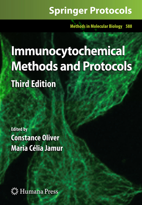
Immunocytochemical Methods and Protocols
Humana Press Inc. (Verlag)
978-1-58829-463-0 (ISBN)
Antibody Preparation.- Overview of Antibodies for Immunochemistry.- to the Purification of Antibodies.- Antibody Purification: Ammonium Sulfate Fractionation or Gel Filtration.- Antibody Purification: Ion-Exchange Chromatography.- Antibody Purification: Affinity Chromatography – Protein A and Protein G Sepharose.- Conjugation of Fluorochromes to Antibodies.- Biotinylation of Antibodies.- Tissue Preparation for Light Microscopic Analysis.- Cell Fixatives for Immunostaining.- Permeabilization of Cell Membranes.- Preparation of Frozen Sections for Analysis.- Processing of Cytological Specimens.- Processing of Tissue Culture Cells.- Processing of Tissue Specimens.- Heat-Induced Antigen Retrieval for Immunohistochemical Reactions in Routinely Processed Paraffin Sections.- Bright Field Detection Systems.- Fluorochromes: Properties and Characteristics.- Direct Immunofluorescent Labeling of Cells.- Fluorescence Labeling of Surface Antigens of Attached or Suspended Tissue-Culture Cells.- Fluorescence Labeling of Intracellular Antigens of Attached or Suspended Tissue-Culture Cells.- Fluorescent Visualization of Macromolecules in Drosophila Whole Mounts.- Overview of Conventional Fluorescence Photomicrography.- Overview of Confocal Microscopy.- Overview of Laser Microbeam Applications as Related to Antibody Targeting.- Immuno-Laser Capture Microdissection of Rat Brain Neurons for Real Time Quantitative PCR.- Overview of Antigen Detection Through Enzymatic Activity.- The Peroxidase–Antiperoxidase (PAP) Method and Other All-Immunologic Detection Methods.- The Avidin–Biotin Complex (ABC) Method and Other Avidin–Biotin Binding Methods.- Avidin-Biotin Labeling of Cellular Antigens in Cryostat-Sectioned Tissue.- Multiple Antigen Immunostaining Procedures.- ImmunoenzymaticQuantitative Analysis of Antigens Expressed on the Cell Surface (Cell-ELISA).- Use of Immunogold with Silver Enhancement.- Fluorescence-Activated Cell Sorter (FACS) Analyses.- Overview of Flow Cytometry and Fluorescent Probes for Flow Cytometry.- Tissue Disaggregation.- Indirect Immunofluorescent Labeling of Viable Cells.- Indirect Immunofluorescent Labeling of Fixed Cells.- Fluorescent Labeling of DNA.- Deparaffinization and Processing of Pathologic Material.- Colloidal Gold Detection Systems for Electron Microscopic Analysis.- Fixation and Embedding.- Preparation of Colloidal Gold.- Conjugation of Colloidal Gold to Proteins.- Colloidal Gold/Streptavidin Methods.- Pre-embedding Labeling Methods.- Postembedding Labeling Methods.- The Clinical Laboratory.- The Clinical Immunohistochemistry Laboratory: Regulations and Troubleshooting Guidelines.
| Reihe/Serie | Methods in Molecular Biology ; 588 |
|---|---|
| Zusatzinfo | 4 Illustrations, color; 40 Illustrations, black and white; XII, 416 p. 44 illus., 4 illus. in color. |
| Verlagsort | Totowa, NJ |
| Sprache | englisch |
| Maße | 178 x 254 mm |
| Themenwelt | Studium ► 2. Studienabschnitt (Klinik) ► Pathologie |
| Studium ► Querschnittsbereiche ► Infektiologie / Immunologie | |
| Naturwissenschaften ► Biologie ► Biochemie | |
| ISBN-10 | 1-58829-463-3 / 1588294633 |
| ISBN-13 | 978-1-58829-463-0 / 9781588294630 |
| Zustand | Neuware |
| Haben Sie eine Frage zum Produkt? |
aus dem Bereich
