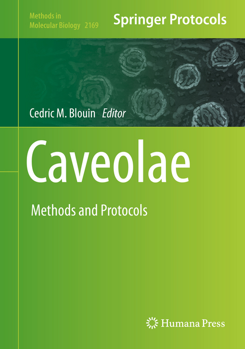
Caveolae
Springer-Verlag New York Inc.
978-1-0716-0731-2 (ISBN)
Authoritative and comprehensive, Caveolae: Methods and Protocols is a valuable resource for both novice and expert researchers who are interested in discovering new roles or regulations of formed caveolae, and proteins composing their coat in various model organisms.
Selective Visualization of Caveolae by TEM using APEX2.- Freeze-Fracture Replica Immunolabeling of Cryopreserved Membrane Compartments, Cultured Cells and Tissues.- Analysis of Caveolin in Primary Cilia.- Method for Efficient Observation of Caveolin-1 Plasma Membrane by Microscopy Imaging Analysis.- Quantitative Image Analysis of the Spatial Organization and Mobility of Caveolin Aggregates at the Plasma Membrane.- Spatiotemporal Analysis of Caveolae Dynamics using Total Internal Reflection Fluorescence Microscopy.- Live-Cell FRET Imaging of Phosphorylation-Dependent Caveolin-1 Switch.- GPMVs as a Tool to Study Caveolin-Interacting Partners.- Biotin Proximity Labeling to Identify Protein-Protein Interactions for Cavin1.- Investigation of Novel Cavin-1/Suppressor of Cytokine 3 (SOCS3) Interactions by Co-Immunoprecipitation, Peptide Pull-Down and Peptide Array Overlay Approaches.- Analysis of Protein and Lipid Interactions using Liposome Co-Sedimentation Assays.- Liposome Binding Assay to Characterize Cavin Family Protein Structure and Functions.- Preparation of Caveolin-1 for NMR Spectroscopy Experiments.- Tagging and Deleting of Endogenous Caveolar Components using CRISPR/Cas9 Technology.- Pulling of Tethers from the Cell Plasma Membrane using Optical Tweezers.- Live Confocal Imaging of Zebrafish Notochord Cells under Mechanical Stress In Vivo.- Study of Caveolae Dependent Mechanoprotection in Huma Muscle Cells using Micropatterning and Live Cell Microscopy.- Immunofluorescence-Based Analysis of Caveolin-3 in the Diagnostic Management of Neuromuscular Diseases.
| Erscheinungsdatum | 30.06.2020 |
|---|---|
| Reihe/Serie | Methods in Molecular Biology ; 2169 |
| Zusatzinfo | 37 Illustrations, color; 9 Illustrations, black and white; XI, 218 p. 46 illus., 37 illus. in color. |
| Verlagsort | New York, NY |
| Sprache | englisch |
| Maße | 178 x 254 mm |
| Themenwelt | Naturwissenschaften ► Biologie ► Biochemie |
| Naturwissenschaften ► Biologie ► Mikrobiologie / Immunologie | |
| Naturwissenschaften ► Biologie ► Zellbiologie | |
| Schlagworte | electron microscopy • endothelial cells • Fluorescence Microscopy • Protein Purification • spinning disk microscopy |
| ISBN-10 | 1-0716-0731-6 / 1071607316 |
| ISBN-13 | 978-1-0716-0731-2 / 9781071607312 |
| Zustand | Neuware |
| Haben Sie eine Frage zum Produkt? |
aus dem Bereich


