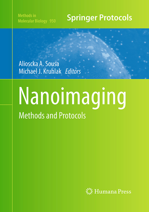
Nanoimaging
Humana Press Inc. (Verlag)
978-1-4939-5961-7 (ISBN)
Authoritative and accessible, Nanoimaging: Methods and Protocols highlights many of the most exciting possibilities in microscopy for the investigation of biological structures at the nano length molecular scales.
Introduction: Nanoimaging Techniques in Biology.- Live-Cell Imaging of Vesicle Trafficking and Divalent Metal Ions by Total Internal Reflection Fluorescence (TIRF) Microscopy.- 4Pi Microscopy.- Fluorescence in situ Hybridization Applications for Super-Resolution 3D Structured Illumination Microscopy.- Two-Color STED Imaging of Synapses in Living Brain Slices.- Super-Resolution Imaging by Localization Microscopy.- High-Content Super-Resolution Imaging of Live Cell by uPAINT.- Super-Resolution Fluorescence Imaging with Blink Microscopy.- Photoswitchable Fluorophores for Single-Molecule Localization Microscopy.- Single-Molecule Tracking of mRNA in Living Cells.- Semiautomatic, High-Throughput, High-Resolution Protocol for Three-Dimensional Reconstruction of Single Particles in Electron Microscopy.- Mass Mapping of Amyloid Fibrils in the Electron Microscope using STEM Imaging.- Elemental Mapping by Electron Energy Loss Spectroscopy in Biology.- Cellular Nanoimaging by Cryo Electron Tomography.- Large-Volume Reconstruction of Brain Tissue from High-Resolution Serial Section Images Acquired by SEM-Based Scanning Transmission Electron Microscopy.- 3D Imaging of Cells and Tissues by Focused Ion Beam/Scanning Electron Microscopy (FIB/SEM).- Preparation of Gold Nanocluster Bioconjugates for Electron Microscopy.- Atomic Force Microscopy Imaging of Macromolecular Complexes.- Imaging of Transmembrane Proteins Directly Incorporated Within Supported Lipid Bilayers Using Atomic Force Microscopy.- Functional AFM Imaging of Cellular Membranes Using Functionalized Tips.- Near-Field Scanning Optical Microscopy for High Resolution Membrane Studies.- Correlative Fluorescence and EFTEM Imaging of the Organized Components of the Mammalian Nucleus.- High Data Output Method for 3-D Correlative Light-Electron Microscopy Using Ultrathin Cryosections.- Correlative Optical and Scanning Probe Microscopies for Mapping Interactions at Membranes.- Nanoimaging Cells Using Soft X-Ray Tomography.- Secondary Ion Mass Spectrometry Imaging of Biological Membranes at High Spatial Resolution.
| Erscheinungsdatum | 19.08.2017 |
|---|---|
| Reihe/Serie | Methods in Molecular Biology ; 950 |
| Zusatzinfo | 76 Illustrations, color; 56 Illustrations, black and white; XIV, 510 p. 132 illus., 76 illus. in color. |
| Verlagsort | Totowa, NJ |
| Sprache | englisch |
| Maße | 178 x 254 mm |
| Themenwelt | Medizinische Fachgebiete ► Radiologie / Bildgebende Verfahren ► Radiologie |
| Medizin / Pharmazie ► Studium | |
| Naturwissenschaften ► Biologie | |
| Naturwissenschaften ► Physik / Astronomie ► Angewandte Physik | |
| ISBN-10 | 1-4939-5961-1 / 1493959611 |
| ISBN-13 | 978-1-4939-5961-7 / 9781493959617 |
| Zustand | Neuware |
| Haben Sie eine Frage zum Produkt? |
aus dem Bereich


