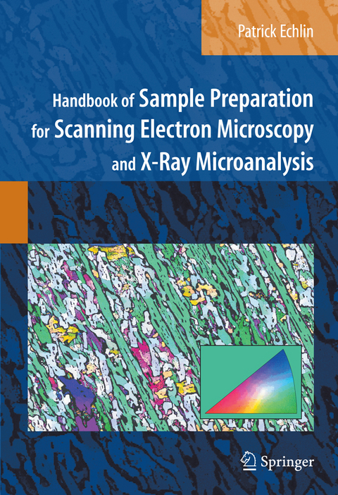
Handbook of Sample Preparation for Scanning Electron Microscopy and X-Ray Microanalysis
Springer-Verlag New York Inc.
978-1-4419-4674-4 (ISBN)
Patrick Echlin was a lecturer in the Department of Plant Sciences and Director of the Multi-Imaging Centre, School of Biological Science, University of Cambridge until he retired in 1999. He has taught for more than thirty years at the Lehigh University Microscopy School and is the author and co-auther of eight books on scanning electron microscopy and x-ray microanalysis. He is an Honorary Fellow of the Royal Microscopical Society and received the Distinguished Scientist Award in Biological Sciences from the Microscope Society of America in 2001.
Sample Collection and Selection.- Sample Preparation Tools.- Sample Support.- Sample Embedding ?and Mounting.- Sample Exposure.- Sample Dehydration.- Sample Stabilization for Imaging in the SEM.- Sample Stabilization to Preserve Chemical Identity.- Sample Cleaning.- Sample Surface Charge Elimination.- Sample Artifacts and Damage.- Additional Sources of Information.
This handbook should find its way to the reference bookshelf of all imaging laboratories. It should also become required reading for anyone being trained for SEM work, or anyone who might need to have their samples examined by using such techniques. In that way, it will be less likely that deficient results will be published and that the full potential of the SEM be realized.
-- Iolo ap Gwynn, Microscopy and Microanalysis (2010)
| Erscheint lt. Verlag | 23.11.2010 |
|---|---|
| Zusatzinfo | 159 Illustrations, color; XII, 332 p. 159 illus. in color. |
| Verlagsort | New York, NY |
| Sprache | englisch |
| Maße | 178 x 254 mm |
| Themenwelt | Naturwissenschaften ► Biologie |
| Naturwissenschaften ► Physik / Astronomie ► Angewandte Physik | |
| Naturwissenschaften ► Physik / Astronomie ► Festkörperphysik | |
| Naturwissenschaften ► Physik / Astronomie ► Thermodynamik | |
| Technik ► Maschinenbau | |
| ISBN-10 | 1-4419-4674-8 / 1441946748 |
| ISBN-13 | 978-1-4419-4674-4 / 9781441946744 |
| Zustand | Neuware |
| Haben Sie eine Frage zum Produkt? |
aus dem Bereich


