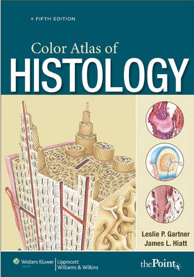
Color Atlas of Histology
Seiten
2009
|
5th revised North American ed
Lippincott Williams and Wilkins (Verlag)
978-0-7817-8872-4 (ISBN)
Lippincott Williams and Wilkins (Verlag)
978-0-7817-8872-4 (ISBN)
- Titel erscheint in neuer Auflage
- Artikel merken
Zu diesem Artikel existiert eine Nachauflage
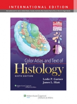
Provides medical, dental, allied health, and biology students with an outstanding collection of histology images for all of the major tissue classes and body systems. This title features a full-color art program comprising over 500 high-quality photomicrographs, scanning electron micrographs, and drawings.
Now in its Fifth Edition, this best-selling atlas provides medical, dental, allied health, and biology students with an outstanding collection of histology images for all of the major tissue classes and body systems. This is a compact lab atlas with relevant concise text and consistent format presentation of photomicrograph plates. With a handy spiral binding that allows ease of use, it features a full-color art program comprising over 500 high-quality photomicrographs, scanning electron micrographs, and drawings. Didactic text at the beginning of each chapter includes an Introduction, Histophysiology, Clinical Correlations, and Overview. A companion Website includes an interactive atlas and a question bank. The interactive atlas contains all the photomicrographs and electron micrographs and accompanying legends from the atlas. Images may be viewed with or without the labels and/or legends, enlarged, or compared side-by-side. A "hotspot" feature allows students to self-test on the labeling.
Now in its Fifth Edition, this best-selling atlas provides medical, dental, allied health, and biology students with an outstanding collection of histology images for all of the major tissue classes and body systems. This is a compact lab atlas with relevant concise text and consistent format presentation of photomicrograph plates. With a handy spiral binding that allows ease of use, it features a full-color art program comprising over 500 high-quality photomicrographs, scanning electron micrographs, and drawings. Didactic text at the beginning of each chapter includes an Introduction, Histophysiology, Clinical Correlations, and Overview. A companion Website includes an interactive atlas and a question bank. The interactive atlas contains all the photomicrographs and electron micrographs and accompanying legends from the atlas. Images may be viewed with or without the labels and/or legends, enlarged, or compared side-by-side. A "hotspot" feature allows students to self-test on the labeling.
1. The Cell 2. Epithelium and Glands 3. Connective Tissue 4. Cartilage and Bone 5. Blood and Hemopoiesis 6. Muscle 7. Nervous Tissue 8. Circulatory System 9. Lymphoid Tissue 10. Endocrine System 11. Integument 12. Respiratory System 13. Digestive System I: Oral Region 14. Digestive System II: Alimentary Canal 15. Digestive System III: Digestive Glands 16. Urinary System 17. Female Reproductive System 18. Male Reproductive System 19. Special Senses
| Erscheint lt. Verlag | 1.2.2009 |
|---|---|
| Zusatzinfo | 580 illustrations |
| Verlagsort | Philadelphia |
| Sprache | englisch |
| Maße | 178 x 254 mm |
| Gewicht | 771 g |
| Themenwelt | Studium ► 1. Studienabschnitt (Vorklinik) ► Histologie / Embryologie |
| ISBN-10 | 0-7817-8872-2 / 0781788722 |
| ISBN-13 | 978-0-7817-8872-4 / 9780781788724 |
| Zustand | Neuware |
| Informationen gemäß Produktsicherheitsverordnung (GPSR) | |
| Haben Sie eine Frage zum Produkt? |
Mehr entdecken
aus dem Bereich
aus dem Bereich
Zytologie, Histologie und mikroskopische Anatomie
Buch | Hardcover (2022)
Urban & Fischer in Elsevier (Verlag)
54,00 €
