MRI Neuroanatomy
Springer International Publishing (Verlag)
978-3-031-76695-4 (ISBN)
- Noch nicht erschienen - erscheint am 14.07.2025
- Versandkostenfrei innerhalb Deutschlands
- Auch auf Rechnung
- Verfügbarkeit in der Filiale vor Ort prüfen
- Artikel merken
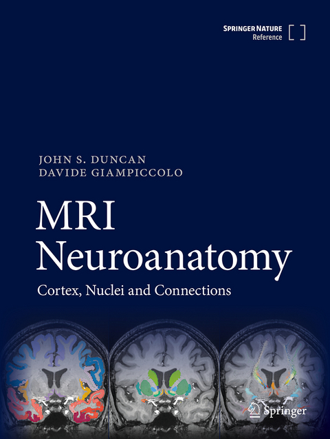
The dogma that brain function relied on the cortex has dominated clinical neurology, neurosurgery and psychiatry for the last 100 years. Since the start of the 2000s, it has become evident that brain function is orchestrated as a network through white matter connections. This framework provides an understanding of brain function and dysfunction, and has radically changed how neurosurgical resections are performed. There is currently no manual for clinicians to visualize this functional anatomy in a fast, easy and user-friendly way. This is particularly important for senior clinicians who may have an understanding of cortical anatomy but may struggle with newly described white matter connections as they may now be visualized with MRI, and also for trainees who are learning the subject of applied neuro-anatomy. With this book, we aim to bridge this gap.
In 1995 Jackson and Duncan produced "MRI Neuroanatomy: a new angle on the brain" using the best clinical MRI available at the time and did not demonstrate white matter tracts. This was very well received, and it is time now to produce an atlas that shows the 3D anatomy of gray and white matter to contemporary standards.
This atlas is based on a high-quality MRI of a healthy subject which resembles the type of imaging is regularly available to clinicians. It is structured in sections (cortical anatomy, subcortical anatomy, and network anatomy) that are intended to guide clinicians from the classical cortical paradigms into a network neuroscience perspective. The 2D orthogonal slices are organized in two orientations:
(A) following the plane of the anterior and posterior commissure, as has been traditionally used in stereotactic atlases
(B) following the plane of the hippocampus, as is commonly used in clinical epilepsy practice.
The first part shows the 3D anatomy of the cerebral hemisphere, deep nuclei, brainstem and cerebellum.
The second part shows the same brain cut in 2D orthogonal slices (axial, coronal, sagittal) with a raw T1-weighted image, accompanied by a labelled image showing the gross anatomy of the brain and grey matter structures and, also, the labelled white matter tracts in that slice. This is particularly relevant for neurosurgeons, who will be able to appreciate before planning a resection the relationship between each tract's trajectory and the gray matter. This will also benefit neurologists, enabling clarity as to how single lesions can cause multiple disconnection and impact on different functions and behaviours.
The third part demonstrates the 3D anatomy of the major white matter tracts in the brain, to indicate how distant lesions can impact the same function.
John Duncan is a Consultant Neurologist specialising in epilepsy, at the National Hospital for Neurology and Neurosurgery, Queen Square, London. His personal research focus is neuroimaging applied to epilepsy surgery.
He was appointed Professor of Neurology at the UCL Institute of Neurology in 1998. In 2004 he received the annual Clinical Research recognition award of the American Epilepsy Society.
He has 52,194 citations, H-index (G) 122. 493 original peer reviewed publications, 88 reviews, 57 chapters and 20 books, including: Jackson GD, Duncan JS. MRI Neuroanatomy: A new angle on the brain. London, Churchill Livingstone, 1996. 303 pp.
Davide Giampiccolo is a complex epilepsy neurosurgical fellow at the National Hospital for Neurology and Neurosurgery and completing a PhD at the UCL Institute of Neurology. He has a keen interest in functional neuroanatomy and connectomics to improve neurological outcome in resective surgery. He trained in white matter anatomy and tractography with Professor Catani, at King's College London. He is expert in intraoperative neurophysiology for network preservation. He was trained in white matter stimulation in Montpellier under Professor Hugues Duffau. With Professor Duffau he recently proposed a reappraisal of current anatomical models for language function (Giampiccolo and Duffau, Brain, 2022). He has authored several papers and book chapters on the topic of cortical and subcortical white matter stimulation aiming to bridge anatomy and electrophysiology. For his studies on asleep language monitoring in 2019 he was the first non-American to receive the annual Academy Award from the American Academy of Neurological Surgery and in 2020 was granted the Integra research fund by the European Association of Neurological Societies.
Introduction to three dimensional anatomy.- 3-dimensional anatomy of the cerebral hemisphere.- 3-dimensional anatomy of the deep nuclei.- 3-dimensional anatomy of the brainstem and cerebellum. 3-dimensional anatomy of brainstem and cerebellar nuclei.- Axial slices of cerebral hemisphere: AC-PC orientation.- Coronal slices of cerebral hemisphere: AC-PC orientation.- Sagittal slices of cerebral hemisphere.- Axial slices of cerebral hemisphere: Hippocampal orientation.- Coronal slices of cerebral hemisphere: Hippocampal orientation.- Axial slices of brainstem and cerebellum: AC-PC orientation.- Coronal slices of brainstem and cerebellum: AC-PC orientation.- Sagittal slices of brainstem and cerebellum.- Introduction to individual tracts in the brain.- Arcuate fasciculus.- Inferior fronto-occipital fasciculus.- Uncinate fasciculus.- Third branch of the superior longitudinal fasciculus.- Second branch of the superior longitudinal fasciculus.- First branch of the superior longitudinal fasciculus.- Posterior segment of the arcuate fasciculus.- Middle longitudinal fasciculus.- Inferior longitudinal fasciculus.- Temporal longitudinal fasciculus.
| Erscheint lt. Verlag | 14.7.2025 |
|---|---|
| Zusatzinfo | XX, 1212 p. 410 illus., 287 illus. in color. |
| Verlagsort | Cham |
| Sprache | englisch |
| Maße | 210 x 279 mm |
| Themenwelt | Medizinische Fachgebiete ► Chirurgie ► Neurochirurgie |
| Medizin / Pharmazie ► Medizinische Fachgebiete ► Neurologie | |
| Schlagworte | Clinical Neuroanatomy • Epilepsy • Medical Imaging • Network neuroscience • neurosurgery • Radiology • tractography • White matter anatomy |
| ISBN-10 | 3-031-76695-4 / 3031766954 |
| ISBN-13 | 978-3-031-76695-4 / 9783031766954 |
| Zustand | Neuware |
| Haben Sie eine Frage zum Produkt? |
aus dem Bereich


